
In vivo models are indispensable for understanding the mechanisms of cardiovascular diseases and for the preclinical testing of new therapeutic agents. These models offer a complex and interactive environment in which the efficacy and safety of pharmacological interventions can be evaluated before proceeding to human trials.
Cardiovascular diseases remain a leading cause of mortality globally, necessitating effective treatments. In vivo models are chosen for their physiological similarities to humans. These models help researchers understand how cardiovascular systems react to potential treatments in a complex organism, closely mirroring human responses.
These models not only allow for testing of drug efficacy and safety but also provide insights into the pathophysiological processes of cardiovascular diseases. This includes the study of molecular pathways, the identification of potential drug targets, and the understanding of disease progression.
Pulmonary Arterial Hypertension (PAH) Disease Models
Pulmonary Arterial Hypertension (PAH) is a severe and progressive disease characterized by high blood pressure in the arteries that supply blood to the lungs. This condition leads to right heart failure and ultimately death if untreated. The complexity of PAH and its pathophysiology make it essential to utilize animal models to study the disease mechanisms, discover potential biomarkers, and develop effective therapies.
Monocrotaline-Induced Rat PAH Model
One of the most commonly used animal models for studying PAH is the Monocrotaline-induced rat model. Monocrotaline (MCT) is a toxic alkaloid derived from the plant Crotalaria spectabilis. When administered to rats, it specifically injures the endothelial cells of the pulmonary arteries, leading to progressive pulmonary hypertension that closely resembles human PAH in several aspects.
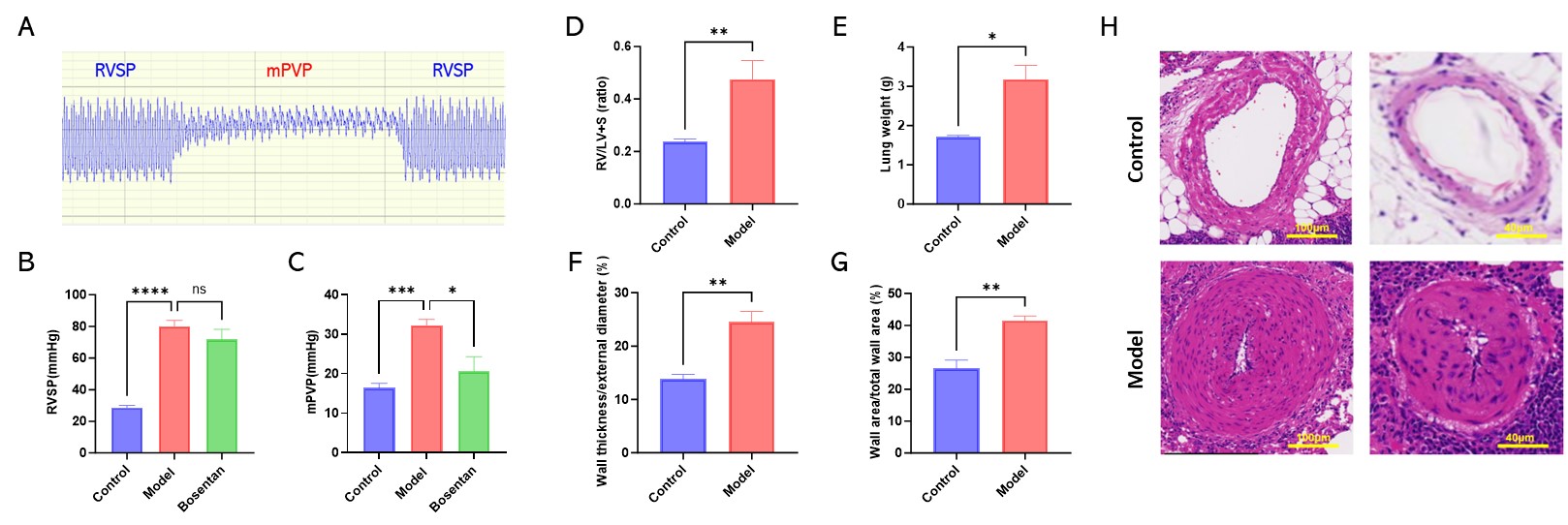
Bosentan alleviates the mPVP in PAH rats. Figure A. Representative image of the RVSP and mPVP. Figure B. The RVSP of PAH rats. Figure C. The mPAP of PAH rats. Figure D. The RV/LV+S ratio in PAH rats. Figure E. The lung weight of PAH rats. Figure F. The wall thickness/external diameter of pulmonary arteries. Figure G. The wall area/total wall area of pulmonary arteries. Figure H. Representative images of the HE staining of pulmonary arteries. Data are presented as mean±SD, n=6 each group.
We value your inquiries and are here to provide you with tailored solutions for your drug discovery and development needs. Whether you have questions, require more information, or are interested in discussing potential collaborations, our team of experts is just a message away.
Feel free to reach out to us.
Address: Bldg 16, Yd 18, Kechuang 13th St, Etown, Tongzhou Dist, Beijing, 100176, China
Email: marketing@ice-biosci.com
Tel:+86-10-67809840