Our kinase activity assays were used in the study of novel FGFR1 inhibitors for triple-negative breast cancer (TNBC).
Abstract
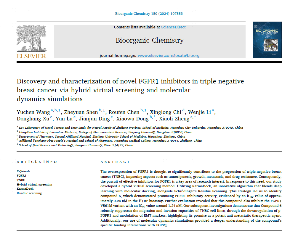
The overexpression of FGFR1 is thought to significantly contribute to the progression of triple-negative breast cancer (TNBC), impacting aspects such as tumorigenesis, growth, metastasis, and drug resistance. Consequently, the pursuit of effective inhibitors for FGFR1 is a key area of research interest. In response to this need, our study developed a hybrid virtual screening method. Utilizing KarmaDock, an innovative algorithm that blends deep learning with molecular docking, alongside Schr¨odinger’s Residue Scanning. This strategy led us to identify compound 6, which demonstrated promising FGFR1 inhibitory activity, evidenced by an IC50 value of approximately 0.24 nM in the HTRF bioassay. Further evaluation revealed that this compound also inhibits the FGFR1 V561M variant with an IC50 value around 1.24 nM. Our subsequent investigations demonstrate that Compound 6 robustly suppresses the migration and invasion capacities of TNBC cell lines, through the downregulation of p- FGFR1 and modulation of EMT markers, highlighting its promise as a potent anti-metastatic therapeutic agent. Additionally, our use of molecular dynamics simulations provided a deeper understanding of the compound’s specific binding interactions with FGFR1.

Our recent collaborative work was published in "Archiv der Pharmazie," demonstrating our excellence in kinase activity assays.
Abstract
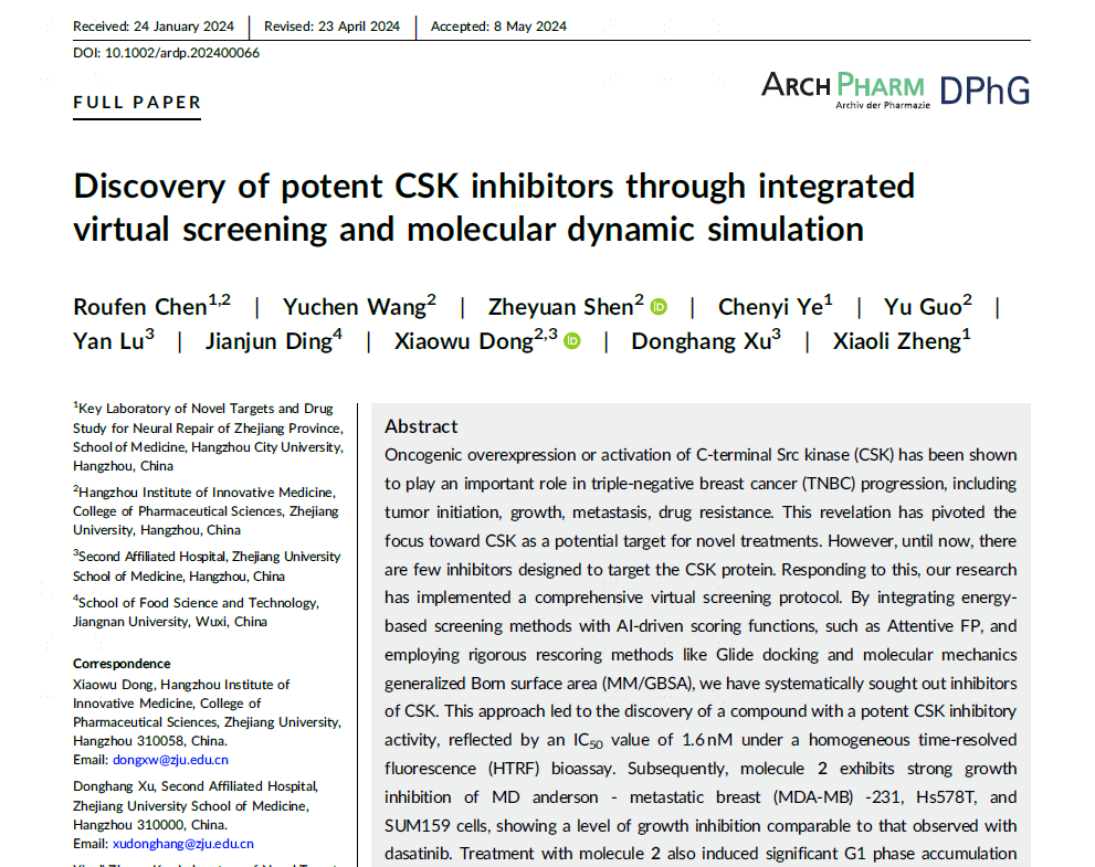
Oncogenic overexpression or activation of C‐terminal Src kinase (CSK) has been shown to play an important role in triple‐negative breast cancer (TNBC) progression, including tumor initiation, growth, metastasis, drug resistance. This revelation has pivoted the focus toward CSK as a potential target for novel treatments. However, until now, there are few inhibitors designed to target the CSK protein. Responding to this, our research has implemented a comprehensive virtual screening protocol. By integrating energybased screening methods with AI‐driven scoring functions, such as Attentive FP, and employing rigorous rescoring methods like Glide docking and molecular mechanics generalized Born surface area (MM/GBSA), we have systematically sought out inhibitors of CSK. This approach led to the discovery of a compound with a potent CSK inhibitory activity, reflected by an IC50 value of 1.6 nM under a homogeneous time‐resolved fluorescence (HTRF) bioassay. Subsequently, molecule 2 exhibits strong growth inhibition of MD anderson ‐ metastatic breast (MDA‐MB) ‐231, Hs578T, and SUM159 cells, showing a level of growth inhibition comparable to that observed with dasatinib. Treatment with molecule 2 also induced significant G1 phase accumulation and cell apoptosis. Furthermore, we have explored the explicit binding interactions of the compound with CSK using molecular dynamics simulations, providing valuable insights into its mechanism of action.

Our Western blot analysis has played a pivotal role in elucidating the effects of the innovative BG-MnGel on the activation of the STING pathway within tumor cells.
Abstract
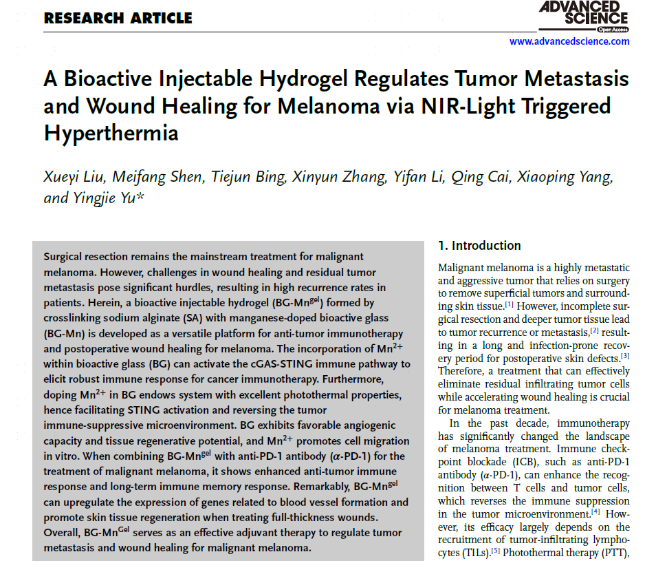
Surgical resection remains the mainstream treatment for malignant melanoma. However, challenges in wound healing and residual tumor metastasis pose significant hurdles, resulting in high recurrence rates in patients. Herein, a bioactive injectable hydrogel (BG-Mngel) formed by crosslinking sodium alginate (SA) with manganese-doped bioactive glass (BG-Mn) is developed as a versatile platform for anti-tumor immunotherapy and postoperative wound healing for melanoma. The incorporation of Mn2+ within bioactive glass (BG) can activate the cGAS-STING immune pathway to elicit robust immune response for cancer immunotherapy. Furthermore, doping Mn2+ in BG endows system with excellent photothermal properties, hence facilitating STING activation and reversing the tumor immune-suppressive microenvironment. BG exhibits favorable angiogenic capacity and tissue regenerative potential, and Mn2+ promotes cell migration in vitro. When combining BG-Mngel with anti-PD-1 antibody (????-PD-1) for the treatment of malignant melanoma, it shows enhanced anti-tumor immune response and long-term immune memory response. Remarkably, BG-Mngel can upregulate the expression of genes related to blood vessel formation and promote skin tissue regeneration when treating full-thickness wounds. Overall, BG-MnGel serves as an effective adjuvant therapy to regulate tumor metastasis and wound healing for malignant melanoma.

Our contribution to the study featured in "Advanced Materials" involves the application of our expertise in flow cytometry analysis, biosafety assessment, and in vivo imaging technologies.
Abstract
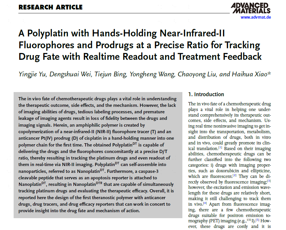
The in vivo fate of chemotherapeutic drugs plays a vital role in understanding the therapeutic outcome, side effects, and the mechanism. However, the lack of imaging abilities of drugs, tedious labeling processes, and premature leakage of imaging agents result in loss of fidelity between the drugs and imaging signals. Herein, an amphiphilic polymer is created by copolymerization of a near-infrared-II (NIR-II) fluorophore tracer (T) and an anticancer Pt(IV) prodrug (D) of cisplatin in a hand-holding manner into one polymer chain for the first time. The obtained PolyplatinDT is capable of delivering the drugs and the fluorophores concomitantly at a precise D/T ratio, thereby resulting in tracking the platinum drugs and even readout of them in real-time via NIR-II imaging. PolyplatinDT can self-assemble into nanoparticles, referred to as NanoplatinDT. Furthermore, a caspase-3 cleavable peptide that serves as an apoptosis reporter is attached to NanoplatinDT, resulting in NanoplatinDTR that are capable of simultaneously tracking platinum drugs and evaluating the therapeutic efficacy. Overall, it is reported here the design of the first theranostic polymer with anticancer drugs, drug tracers, and drug efficacy reporters that can work in concert to provide insight into the drug fate and mechanism of action.