In Vitro Bystander Effect Assays for ADCs are designed to evaluate how the cytotoxic payload released by an ADC can affect not only the target antigen-positive (Ag+) cancer cells but also the neighboring antigen-negative (Ag-) cells. This bystander effect occurs when the payload, once released from the ADC within a targeted cancer cell, diffuses into the surrounding tumor microenvironment, potentially killing nearby non-target cells that do not express the target antigen.
Utilizing advanced cell culture techniques and cell-based assays, this assay provides critical insights into the ability of ADCs to release cytotoxic agents that diffuse and kill nearby antigen-negative cells, thereby enhancing overall therapeutic efficacy.
☑️ Holistic Efficacy Understanding: We assess the impact of ADCs on both target and surrounding tumor cells, addressing tumor heterogeneity.
☑️ Real-Time Precision: Our methods provide real-time, quantitative insights into payload diffusion and impact, optimizing ADC formulations.
☑️ Streamlined, Ready-to-Use Models: Our engineered cell lines and in vivo models accelerate bystander effect evaluation with high reproducibility.
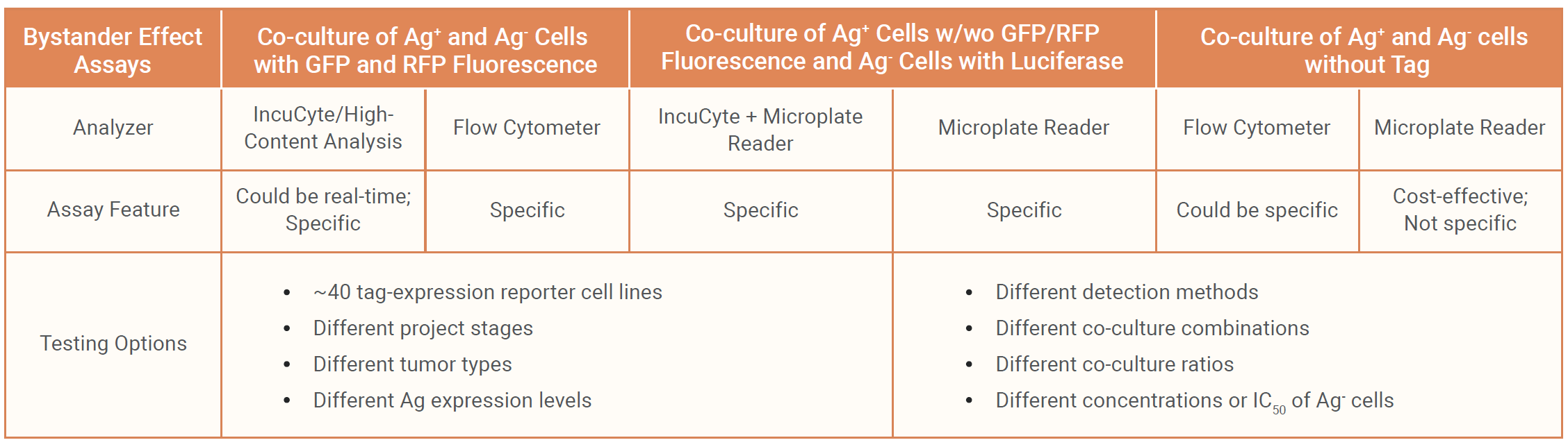
Our assays provide real-time, live-cell imaging to evaluate the bystander effect of ADCs. This service allows continuous monitoring of both antigen-positive and antigen-negative cells in co-culture, tracking how the released payload affects neighboring cells over time. Contact us for more detailed reference data.
Our Flow Cytometry-Based Bystander Effect Assays offer a precise and high-throughput method to evaluate the impact of ADCs on both target (antigen-positive) and non-target (antigen-negative) cells. By using flow cytometry, we can quantify cell viability, apoptosis, and other markers in co-cultured cells, providing detailed insights into how the released payload affects neighboring cells. Contact us for more detailed reference data.
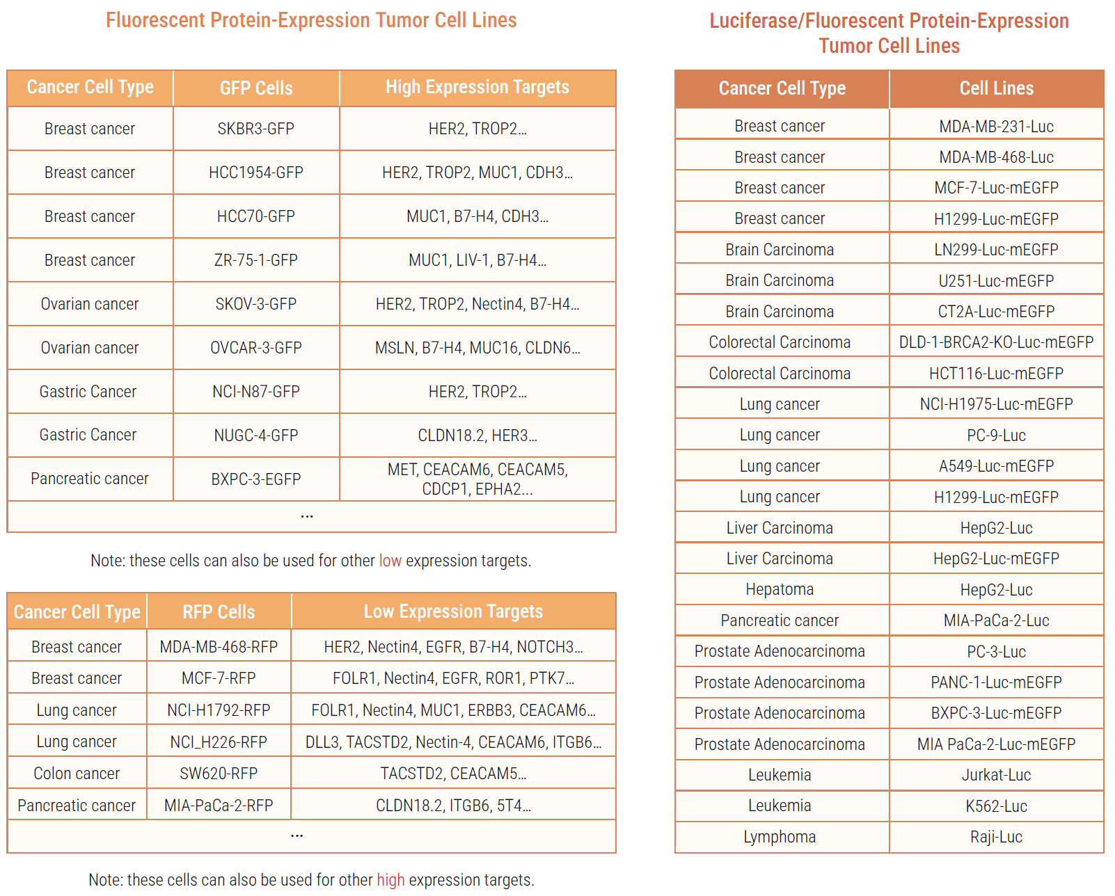
Our Tag-expression Reporter Tumor Cell Lines are engineered to express fluorescent or luminescent tags, enabling specifically monitoring of ADC interactions and effects. These cell lines simplify and accelerate ADC research by allowing precise tracking of efficacy in target and bystander cells. With these reporter lines, researchers can efficiently evaluate ADC bystander effect, optimize linker-payload design, and understand the mechanisms of action, enhancing the development of targeted cancer therapies. Contact us for more detailed reference data.
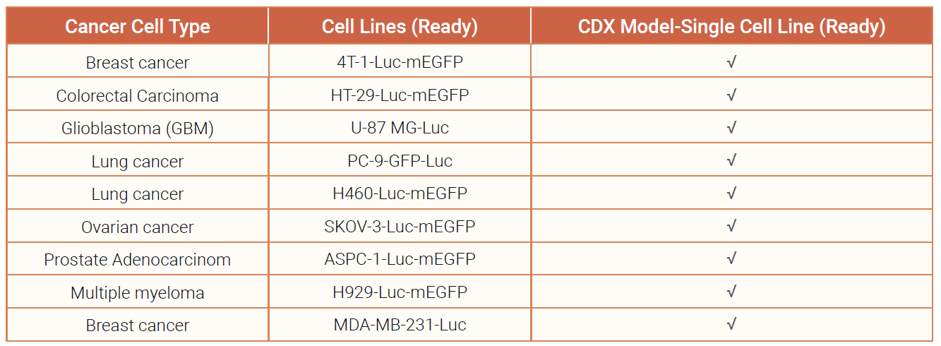
The in vivo bystander effect refers to the phenomenon where cytotoxic agents released by targeted cancer cells (those directly affected by ADCs) can diffuse and induce cell death in neighboring non-targeted cancer cells within a living organism. By using tumor cell lines engineered to express luciferase, we can visualize and monitor the bystander effect using our IVIS in vivo imaging systems.
Using untagged Ag+ cells and luciferase-tagged Ag- cells in co-inoculation CDX models is an effective approach to evaluate the in vivo bystander effect. The luciferase tag on Ag- cells allows for precise and specific monitoring of their response to the ADC treatment, ensuring that the effects observed are due to the bystander effect mediated by the Ag+ cells. This method provides clear and valuable insights into the efficacy of ADCs in heterogeneous tumor environments. Contact us for more detailed reference data.
We value your inquiries and are here to provide you with tailored solutions for your drug discovery and development needs. Whether you have questions, require more information, or are interested in discussing potential collaborations, our team of experts is just a message away.
Feel free to reach out to us.
Address: Bldg 16, Yd 18, Kechuang 13th St, Etown, Tongzhou Dist, Beijing, 100176, China
Email: marketing@ice-biosci.com
Tel:+86-10-67809840