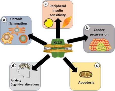
Voltage-gated potassium channels (Kv) were discovered in human and mouse T cells in 1984, and the gene encoding this channel was identified as Kv1.3/KCNA3 in 1990. Each Kv1.3 subunit contains six transmembrane domains (S1-S6), a pore, and intracellular N- and C-termini. Four subunits form a functional tetramer. Kv1.3 channels are primarily distributed in lymphocytes (T and B lymphocytes), the central nervous system, and other tissues and organs. They play significant roles in various physiological and pathological processes, such as neurotransmitter release, insulin secretion, and neuronal excitability.
Impaired Kv1.3 expression can alter insulin sensitivity in adipocytes and skeletal muscle. Most evidence supports that Kv1.3 expression is associated with tumor behavior and malignancy. Kv1.3 is present in the inner membrane of mitochondria, and increased activity is related to the onset and sensitivity of apoptosis. Abnormal activity of Kv1.3 in the central nervous system during insulin resistance is linked to cognitive changes and the development of anxiety. Increased expression of Kv1.3 in activated immune cells, such as macrophages, dendritic cells, and T cells, can lead to chronic and degenerative inflammatory diseases [1]
doi.org/10.1080/14728222.2017.1420170 [1]
Kv1.3 Channels and Autoimmune Diseases
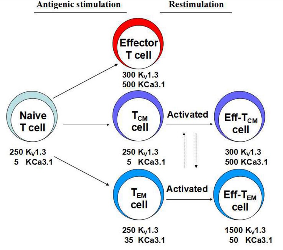
Kv1.3 plays a crucial role in the activation of T lymphocytes, particularly in effector memory T cells (TEM) [2]. When T lymphocytes are activated by antigen-presenting cells (APCs), the intracellular Ca2+ concentration increases, leading to membrane depolarization and the activation of two potassium channels: Kv1.3 and KCa3.1. Kv1.3 promotes the calcium homeostasis required for T cell receptor-mediated activation, gene transcription, and proliferation. In resting T cells, each cell expresses 400 Kv1.3 channels and 5-35 KCa3.1 channels. Upon T cell activation, KCa3.1 expression is upregulated, while Kv1.3 expression remains relatively unchanged. However, after multiple rounds of antigen stimulation, Kv1.3 expression is upregulated, with minimal changes in KCa3.1 expression. This phenomenon is also observed in rat T cells.
Although both Kv1.3 and KCa3.1 contribute to maintaining the driving force for Ca2+ influx, their expression differs. KCa3.1 is highly expressed in central memory T cells (TCM), whereas Kv1.3 is highly expressed in effector memory T cells (TEM). Effector memory T cells are associated with the onset of autoimmune diseases. Blocking Kv1.3 in effector memory T cells can inhibit T cell depolarization, calcium signaling, and cytokine production. Therefore, selective Kv1.3 blockers are considered potential new therapies for treating autoimmune diseases such as multiple sclerosis, type 1 diabetes, psoriasis, rheumatoid arthritis, transplant rejection, Sjögren’s syndrome, and systemic lupus erythematosus [3].
doi:10.3390/toxins7051749 [3]
The Relationship Between Kv1.3 and T Cell Activation
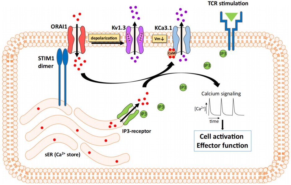
When a specific antigen (green triangle) stimulates the T cell receptor (TCR), it activates the PLC pathway, increasing the concentration of IP3. This increase in IP3 activates the calcium stores in the endoplasmic reticulum (ER), causing the release of Ca2+ into the cytoplasm. The sharp decline in calcium concentration within the ER then triggers the calcium sensor STIM1 to open the CRAC channels, allowing calcium ions to flow in. The continuous influx of calcium ions depolarizes the cell membrane, activating the Kv1.3 and KCa3.1 potassium channels, which leads to the efflux of potassium ions and subsequent membrane repolarization. This process maintains the strong driving force for calcium entry required for T cell activation and proliferation. The elevated intracellular calcium concentration promotes T cell activation and proliferation. Blocking the Kv1.3 channel reduces potassium ion efflux, keeping the membrane depolarized, which in turn reduces Ca2+ entry through the CRAC channels, thereby inhibiting T cell activity [4].
doi.org/10.1007/s42977-021-00071-7 [7]
ICE Bioscience has developed comprehensive screening services for drugs targeting the Kv1.3 channel, utilizing both manual and automated patch clamp techniques. We also support subtype selectivity and use-dependent studies. In this study, we used several reference compounds for Kv1.3 and obtained various validation data.
1. Psora-4
Psora-4 is an effective and selective Kv1.3 inhibitor with an IC50 of 3 nM [6]. Psora-4 demonstrates immunosuppressive properties by inhibiting the in vitro proliferation of myelin-specific effector memory T cells in both humans and rats. The electrophysiology team at ICE Bioscience validated the subtype selectivity of Psora-4. As shown in the figure below, Psora-4 demonstrated much higher selectivity for Kv1.3 over other Kv1-family channels.

Then, we tested the inhibitory effect of Psora-4 on Kv1.3 channel currents using both manual and automated patch clamp techniques. The IC50 values obtained were 1.362 nM and 6.006 nM, respectively.
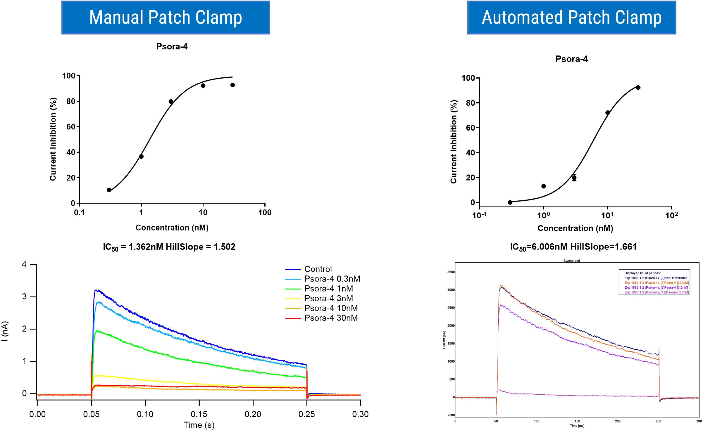
Using the stimulation protocol provided in reference 6, we confirmed that Psora-4 preferentially binds to the C-type inactivated state of the Kv1.3 channel and blocks Kv1.3 in a use-dependent manner.
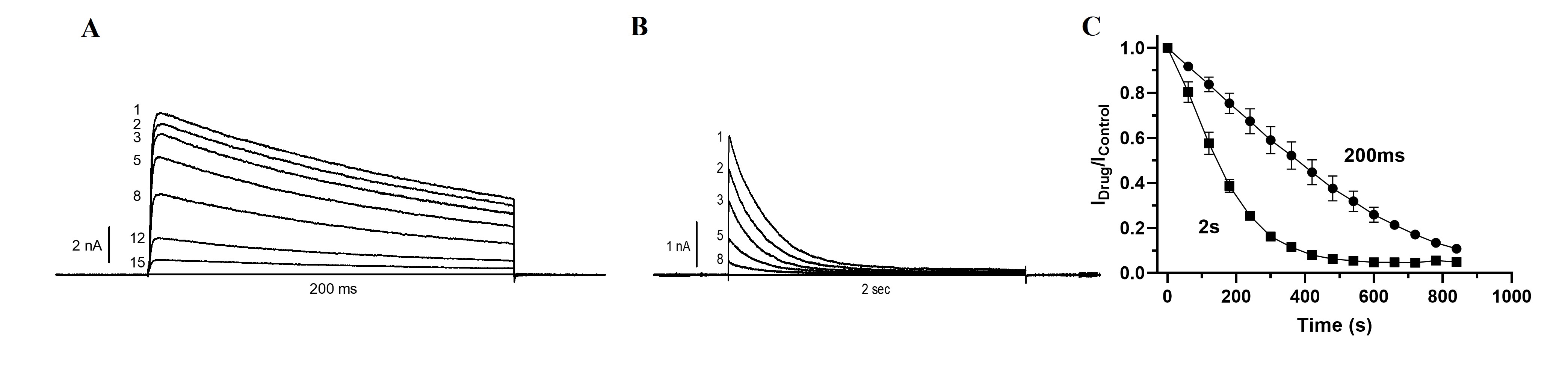
Figure 1. The Kv1.3 control current was induced by a 200-ms depolarizing pulse to +40 mV. Psora-4 (10 nM) was then introduced to the bath while maintaining the membrane potential at -80 mV. After 5 minutes, consecutive 200-ms or 2-second pulses were applied every 60 seconds. (A and B) Show the inhibition of Kv1.3 following the application of 10 nM Psora-4 during consecutive 200-ms and 2-second pulses (pulses 1–15). (C) Illustrates the effect of varying pulse duration on the time required to reach a steady-state block.
2. PAP-1
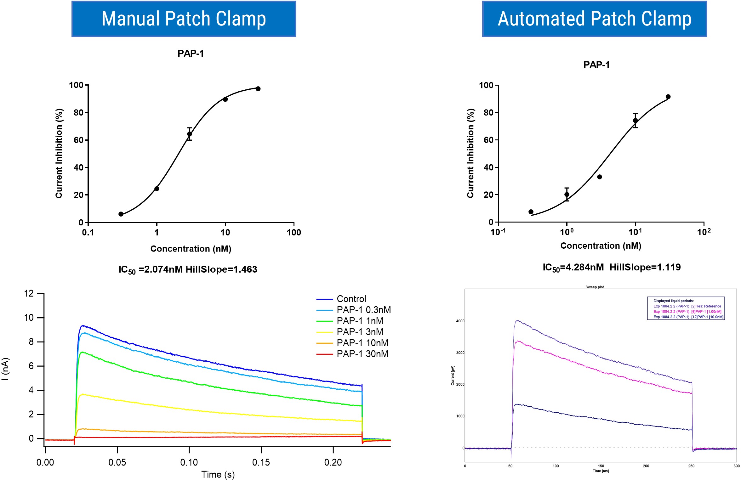
PAP-1 (5-(4-Phenoxybutoxy)psoralen) is a potent, selective, oral Kv1.3 channel blocker with an IC50 of 2 nM [5]. PAP-1 blocks Kv1.3 in a use-dependent manner by preferentially binding to the C-type inactivated state of the channel. It exhibits 23-fold selectivity over Kv1.5 and 33- to 125-fold selectivity over all other Kv1.X family channels. Employing both manual and automated patch clamp techniques, we confirmed the inhibitory effect of PAP-1 on Kv1.3 channel currents, with IC50 values of 2.074 nM and 4.284 nM, respectively.
3. ShK & Margatoxin
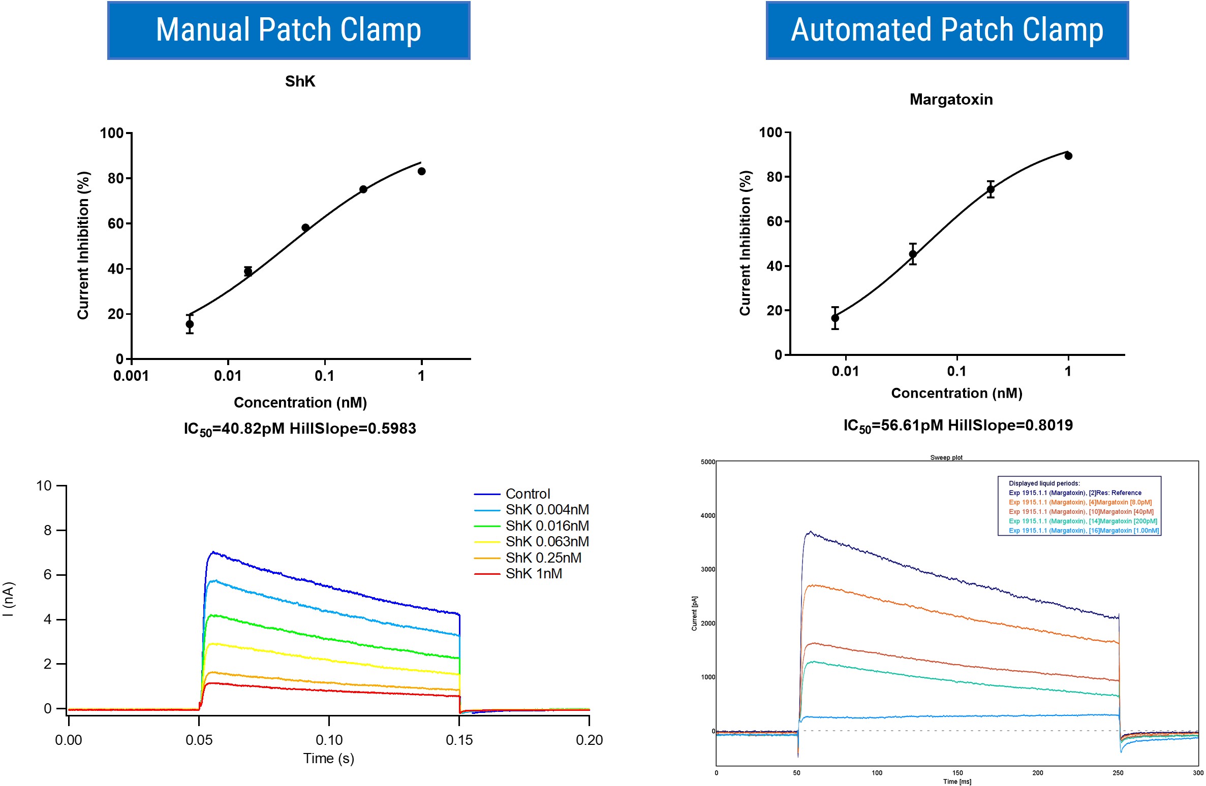
Kv1.3 inhibitors can be broadly classified into three categories: small molecule compounds, animal toxins, and defensins. Animal toxins, in comparison to small molecules, offer more precise and potent mechanisms of action. They are also widely available and abundant, constituting a rich source for peptide drug development. Notably, ShK (Stichodactyla helianthus Neurotoxin) is a specific peptide inhibitor of the Kv1.3 potassium channel, while Margatoxin, an α-KTx scorpion toxin, exhibits high-affinity inhibition of Kv1.3. The electrophysiology team at ICE Bioscience has also confirmed the effects of these two peptide compounds on Kv1.3 channel currents.
In summary, our comprehensive assay results are consistent with those reported in the literature. We also offer clients a variety of other reference compounds targeting Kv1.3. Furthermore, our well-established stable cell lines facilitate the assessment of drug efficacy on Kv1, Kv2, Kv3, Kv4, and Kv7 channel families.
References
[1] Serrano-Albarras, A., et al., Kv1.3: a multifunctional channel with many pathological implications. Expert Opin Ther Targets, 2018. 22(2): p. 101-105.
[2] Hu, L., et al., Characterization of the functional properties of the voltage-gated potassium channel Kv1.3 in human CD4+ T lymphocytes. J Immunol, 2007. 179(7): p. 4563-70.
[3] Zhao, Y., et al., Toxins Targeting the Kv1.3 Channel: Potential Immunomodulators for Autoimmune Diseases. Toxins (Basel), 2015. 7(5): p. 1749-64.
[4] Beeton, C., et al., Kv1.3 channels are a therapeutic target for T cell-mediated autoimmune diseases. Proc Natl Acad Sci U S A, 2006. 103(46): p. 17414-9.
[5] Schmitz, A., et al., Design of PAP-1, a selective small molecule Kv1.3 blocker, for the suppression of effector memory T cells in autoimmune diseases. Mol Pharmacol, 2005. 68(5): p. 1254-70.
[6] Vennekamp, J., et al., Kv1.3-blocking 5-phenylalkoxypsoralens: a new class of immunomodulators. Mol Pharmacol, 2004. 65(6): p. 1364-74.
[7] Varga, Z., Tajti, G., Panyi, G. The Kv1.3 K+ channel in the immune system and its “precision pharmacology” using peptide toxins. BIOLOGIA FUTURA, 2021. 72: p. 75–83.
2026-07-03
2026-05-22
2026-05-21
2026-05-13
We value your inquiries and are here to provide you with tailored solutions for your drug discovery and development needs. Whether you have questions, require more information, or are interested in discussing potential collaborations, our team of experts is just a message away.
Feel free to reach out to us.

Address: Bldg 16, Yd 18, Kechuang 13th St, Etown, Tongzhou Dist, Beijing, 100176, China
Email: marketing@ice-biosci.com
Tel:+86-10-67809840


Get a quote

Go top