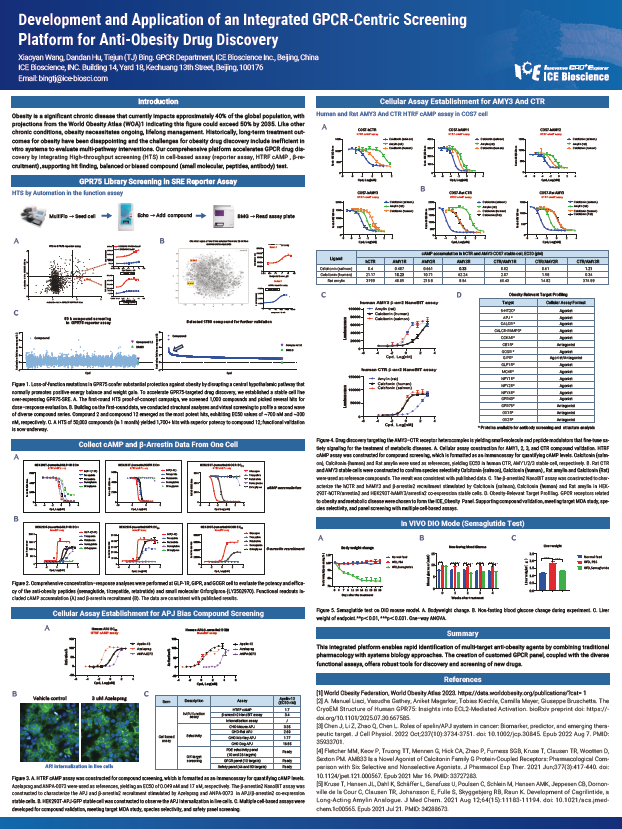
Obesity is a significant chronic disease that currently impacts approximately 40% of the global population, with projections from the World Obesity Atlas (WOA)1 indicating this figure could exceed 50% by 2035. Like other chronic conditions, obesity necessitates ongoing, lifelong management. Historically, long-term treatment out-comes for obesity have been disappointing and the challenges for obesity drug discovery include inefficient in vitro systems to evaluate multi-pathway interventions. Our comprehensive platform accelerates GPCR drug discovery by integrating High-throughput screening (HTS) in cell-based assay (reporter assay, HTRF cAMP, β-recruitment), supporting hit finding, balanced or biased compound (small molecular, peptides, antibody) test.

Targeted protein degradation (TPD) harnesses the ubiquitin-proteasome system to eliminate disease drivers once deemed “undruggable”. PROTACs recruit E3 ligases to targets via two linked warheads, while monovalent molecular glue degraders (MGDs) induce novel ligase-substrate interactions and ternary complex formation. Preclinical success in targeting VAV1, STAT6, NEK7, IRAK4 etc. has demonstrated remarkable efficacy in rheumatoid arthritis, atopic dermatitis and other autoimmune models, positioning TPD as a transformative therapeutic modality.
ICE’s integrated platform accelerates TPD discovery through SpS-based high-throughput screening, targeted libraries and an end-to-end workflow from hit identification to in vivo validation. Our experience includes support for discovery of multiple MGDs and PROTACs—showcased here by discovery of a potential NEK7 MGD, and our integrated TPD platform including orthogonal biochemical and biophysics binding assays, comprehensive in vitro functional characterization, in depth MOA studies, in vivo model validation and safety evaluation. By delivering robust, actionable data at every stage, ICE empowers rapid advancement of next-generation autoimmune interventions.
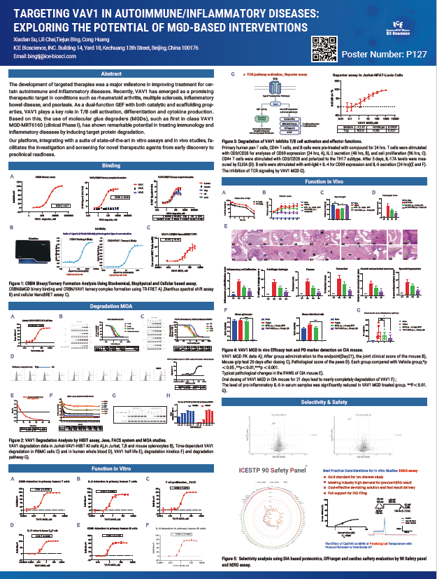
The development of targeted therapies was a major milestone in improving treatment for certain autoimmune and inflammatory diseases. Recently, VAV1 has emerged as a promising therapeutic target in conditions such as rheumatoid arthritis, multiple sclerosis, inflammatory bowel disease, and psoriasis. As a dual-function GEF with both catalytic and scaffolding properties, VAV1 plays a key role in T/B cell activation, differentiation and cytokine production. Based on this, the use of molecular glue degraders (MGDs), such as first in class VAV1 MGD-MRT6160 (clinical Phase I), has shown remarkable potential in treating immunology and inflammatory diseases by inducing target protein degradation.
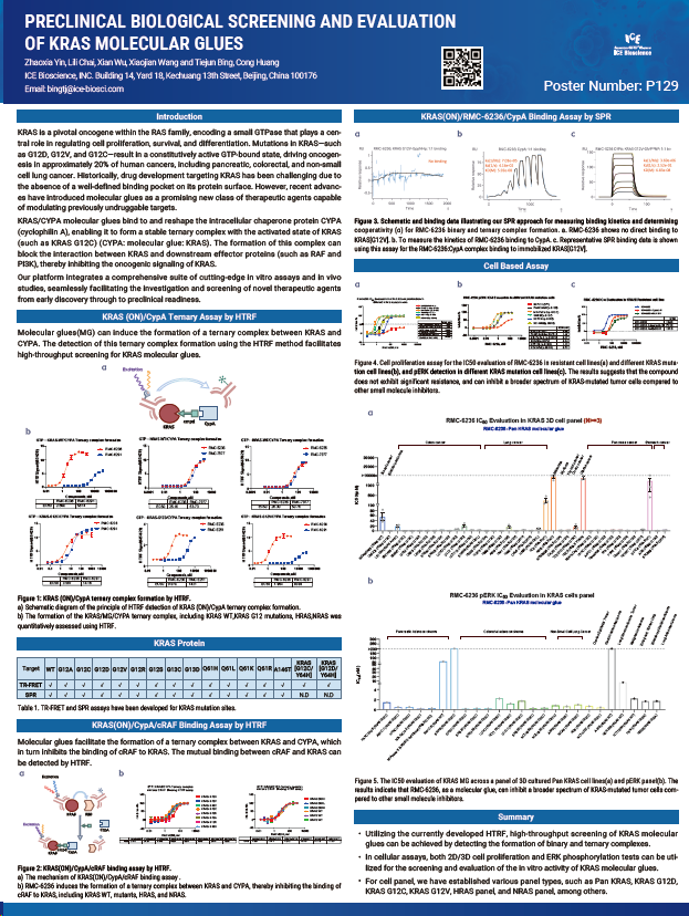
KRAS is a pivotal oncogene within the RAS family, encoding a small GTPase that plays a central role in regulating cell proliferation, survival, and differentiation. Mutations in KRAS—such as G12D, G12V, and G12—result in a constitutively active GTP-bound state, driving oncogenesis in approximately 20% of human cancers, including pancreatic, colorectal, and non-small cell lung cancer. Historically, drug development targeting KRAS has been challenging due to the absence of a well-defined binding pocket on its protein surface. However, recent advances have introduced molecular glues as a promising new class of therapeutic agents capable of modulating previously undruggable targets.
Address: Bldg 16, Yd 18, Kechuang 13th St, Etown, Tongzhou Dist, Beijing, 100176, China
Email: marketing@ice-biosci.com
Tel:+86-10-67809840