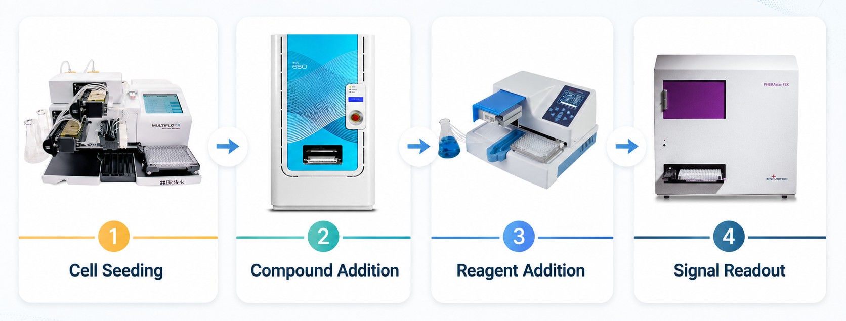
ADC and payload-resistant models can be used for more than confirming loss of response to the original drug. By comparing parental and resistant cells side by side, they can also reveal two opposite response patterns: compounds that lose activity in resistant cells and compounds that become more active in the resistant state.
In this case study, 2,721 compounds from an approved drug library were screened at 100 nM for 7 days in parental HCC1806 cells and three resistant derivatives: HCC1806/Dxd R, HCC1806/SN-38 R, and HCC1806/IMMU-132 R.
The key analytical step was to classify each compound by differential inhibition between parental and resistant cells. Compounds with large activity loss in resistant cells were used to define cross-resistance patterns, while resistant-cell sensitive compounds were advanced as potential vulnerability candidates for dose-response and combination testing.
The screen used one parental model and three related resistant models. HCC1806/Dxd R and HCC1806/SN-38 R represent free payload-resistant contexts linked to topoisomerase I inhibitor biology. HCC1806/IMMU-132 R represents resistance after exposure to a TROP2-directed ADC carrying an SN-38 payload.
This matched design allowed the same compound to be evaluated across parental and resistant backgrounds, making it possible to separate general cytotoxicity from resistance-associated loss or gain of activity.
The full approved-drug library covers approximately 3,200 approved drugs across annotated pharmacological categories, including apoptosis, autophagy, antibacterial agents, receptor modulators, ion-channel modulators, kinase inhibitors, topoisomerase inhibitors, and DNA damage response-related compounds.
In this HCC1806 workflow, the screen used 2,721 compounds available in DMSO. The annotation was useful for interpreting validated hits, because sensitivity or resistance shifts could be linked back to known drug classes instead of being treated as anonymous cytotoxic signals.

Primary screening results were classified using percent inhibition in parental and resistant cells. RS indicated resistant-cell sensitivity, defined as less than 50% inhibition in parental cells and at least 50% inhibition in resistant cells. BI indicated both-cell insensitivity, defined as less than 50% inhibition in both parental and resistant cells.
The Δ% inhibition group captured the opposite pattern. It was calculated as the difference between parental-cell inhibition and resistant-cell inhibition. For example, if parental cells showed 60% inhibition and resistant cells showed 10% inhibition, the Δ value was 50%. A larger Δ therefore indicated a stronger loss of activity in resistant cells and a higher likelihood of differential resistance.
This distinction is important because RS and Δ represent opposite biological directions. RS identifies compounds that may reveal acquired resistant-cell vulnerabilities, while Δ identifies compounds whose activity is reduced in resistant cells and therefore helps define cross-resistance or multi-drug resistance patterns.
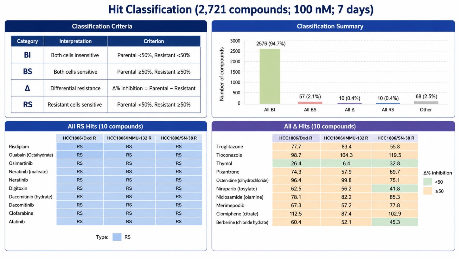
Most compounds were inactive under the primary condition: 2,576 compounds, or 94.7% of the screened set, were classified as all-BI. Only 57 compounds, or 2.1%, were broadly active across parental and resistant models.
The differential-response groups were much smaller. 10 compounds showed an all-RS pattern across the three resistant models, indicating consistent resistant-cell sensitivity. Another 10 compounds showed an all-Δ pattern, indicating consistent activity loss in resistant cells. The remaining 68 compounds showed mixed differential responses across the three resistant models.
Together, the 10 all-RS, 10 all-Δ, and 68 mixed-response compounds formed an 88-compound candidate set for dose-response validation.
Dose-response validation identified 10 multi-drug resistance compounds. These compounds showed reduced activity in at least one resistant model and were therefore used to define resistance-associated drug response patterns, not as resistance-overcoming candidates.
A more informative pattern emerged when the compounds were interpreted by mechanism rather than by RI values alone. Dxd and SN-38 are both linked to topoisomerase I inhibitor biology. In this validation set, the most consistent shared resistance signals included Belotecan hydrochloride and Topotecan hydrochloride, two topoisomerase I inhibitors, as well as Niraparib, a PARP inhibitor connected to DNA damage response biology.
The SN-38-resistant model showed resistance to Belotecan and Topotecan, but not to several topoisomerase II- or anthracycline-like agents such as Epirubicin, Pixantrone, and Teniposide. This suggests that the SN-38 R phenotype was not a broad resistance state against all DNA-damaging agents, but was more consistent with a Topo I-enriched resistance pattern.
By contrast, the Dxd-resistant model showed RI > 5 for a broader set of compounds, including Topo I inhibitors, PARP-related biology, and several Topo II- or anthracycline-like agents. This broader profile suggests that HCC1806/Dxd R may have acquired a wider cytotoxic-stress resistance state than HCC1806/SN-38 R. The IMMU-132-resistant model showed the narrowest pattern, with RI > 5 mainly retained for Belotecan, Niraparib, and Topotecan, indicating that ADC-induced resistance did not fully phenocopy free payload-induced resistance.
| Compound | HCC1806/Dxd R RI | HCC1806/SN-38 R RI | HCC1806/IMMU-132 R RI | Annotated Target / Mechanism | Resistance Pattern |
|---|---|---|---|---|---|
| Shared resistance signals across all three resistant models | |||||
| Belotecan hydrochloride | 14.45 | 7.00 | 8.34 | Topoisomerase | Shared resistance |
| Niraparib | 13.57 | 10.61 | 12.69 | Apoptosis; PARP | Shared resistance |
| Topotecan hydrochloride | 22.16 | 21.11 | 17.75 | Apoptosis; Autophagy; Topoisomerase | Shared resistance |
| Model-selective resistance signals | |||||
| Epirubicin hydrochloride | 7.77 | 1.84 | 1.26 | Antibiotic; Apoptosis; DNA/RNA Synthesis; Topoisomerase | Dxd R-enriched |
| Fumagillin | 92.36 | 7.55 | 2.17 | Antibiotic; HIV; Parasite | Dxd R / SN-38 R-enriched |
| Merimepodib | 2.57 | 11.02 | 4.36 | Dengue virus; Flavivirus; HBV; HCV | SN-38 R-enriched |
| Omaveloxolone | 1.20 | 6.81 | 0.44 | Apoptosis; Keap1-Nrf2; STING | SN-38 R-enriched |
| Pixantrone | 7.89 | 2.16 | 1.52 | Topoisomerase | Dxd R-enriched |
| Teniposide | 5.17 | 0.68 | 0.78 | Topoisomerase | Dxd R-enriched |
| Valrubicin | 5.89 | 2.37 | 1.77 | Antibiotic; PKC | Dxd R-enriched |
Dose-response validation identified 14 multi-drug sensitivity candidates. Unlike the resistance table, the sensitivity hits were strongly enriched in signaling-pathway inhibitors, especially EGFR/ERBB-family inhibitors and MEK inhibitors.
The most striking pattern was observed in HCC1806/SN-38 R. Nearly all 14 candidates showed RI values below 0.2 in the SN-38-resistant model, including EGFR/ERBB inhibitors such as Afatinib, Almonertinib, Dacomitinib, Erlotinib, Icotinib, Lapatinib, Mobocertinib, Neratinib, Osimertinib, and Simotinib, as well as MEK inhibitors such as Cobimetinib and Trametinib.
This suggests that the SN-38-resistant state may have acquired a collateral vulnerability to receptor tyrosine kinase and MAPK-pathway inhibition. The pattern was less broad in HCC1806/Dxd R and narrowest in HCC1806/IMMU-132 R, where Ibrutinib was the only candidate with RI below 0.2.
This model-dependent distribution is important: free SN-38 resistance, free Dxd resistance, and IMMU-132 ADC resistance did not produce identical secondary vulnerabilities, even though all three models are connected to topoisomerase I payload-related resistance.
| Compound | HCC1806/Dxd R RI | HCC1806/SN-38 R RI | HCC1806/IMMU-132 R RI | Annotated Target / Mechanism | Sensitivity Pattern |
|---|---|---|---|---|---|
| EGFR/ERBB-family inhibitor sensitivity enriched in HCC1806/SN-38 R | |||||
| Afatinib | 0.40 | 0.14 | 0.69 | Akt; Apoptosis; Autophagy; c-Met/HGFR; EGFR; p38 MAPK | SN-38 R-enriched |
| Almonertinib | 0.50 | 0.17 | 0.87 | EGFR | SN-38 R-enriched |
| Dacomitinib | 0.10 | 0.04 | 0.25 | Apoptosis; EGFR | Dxd R / SN-38 R-enriched |
| Erlotinib hydrochloride | 0.19 | 0.05 | 0.64 | Autophagy; EGFR | Dxd R / SN-38 R-enriched |
| Icotinib | 0.10 | 0.03 | 0.27 | EGFR | Dxd R / SN-38 R-enriched |
| Lapatinib ditosylate | 0.22 | 0.08 | 0.86 | Autophagy; EGFR; Ferroptosis | SN-38 R-enriched |
| Mobocertinib | 0.35 | 0.12 | 0.86 | EGFR | SN-38 R-enriched |
| Neratinib maleate | 0.12 | 0.07 | 0.43 | EGFR | Dxd R / SN-38 R-enriched |
| Osimertinib | 0.20 | 0.10 | 0.44 | EGFR | SN-38 R-enriched |
| Simotinib | 0.10 | 0.02 | 0.29 | EGFR | Dxd R / SN-38 R-enriched |
| MEK / MAPK-pathway inhibitor sensitivity enriched in HCC1806/SN-38 R | |||||
| Cobimetinib hemifumarate | 0.27 | 0.05 | 2.36 | Apoptosis; MEK | SN-38 R-enriched |
| Trametinib | 0.22 | 0.01 | 0.38 | Apoptosis; Autophagy; MEK | SN-38 R-enriched |
| Other sensitivity candidates | |||||
| Ibrutinib | 0.08 | 0.02 | 0.18 | BTK; Ligands for Target Protein for PROTAC | Shared sensitivity |
| Lonafarnib | 0.03 | 0.33 | 0.22 | Autophagy; Farnesyl Transferase; Ras | Dxd R-enriched |
The 14 multi-drug sensitivity candidates were then evaluated as Drug B in 6×6 matrix combinations with the original ADC or payload as Drug A. The tested Drug A conditions included SN-38, IMMU-132, and Dxd in matched parental and resistant HCC1806 models.
| Cell Model | Drug A | Drug B | Matrix Format |
|---|---|---|---|
| HCC1806 | SN-38 | 14 multi-drug sensitivity candidates | 6 × 6 matrix |
| HCC1806 | IMMU-132 | ||
| HCC1806 | Dxd | ||
| HCC1806/SN-38 R | SN-38 | ||
| HCC1806/IMMU-132 R | IMMU-132 | ||
| HCC1806/Dxd R | Dxd |
The most relevant resistant-model signal was IMMU-132 + Lapatinib in HCC1806/IMMU-132 R cells, which reached the synergy threshold by both Bliss and HSA scoring. This result is biologically informative because Lapatinib was not a strong single-agent sensitivity hit in the IMMU-132-resistant model, suggesting that its value emerged mainly in the ADC combination context.
One possible interpretation is that ERBB-family signaling may help buffer the resistant cells against ADC/payload-induced stress. IMMU-132 delivers an SN-38 payload, which is linked to topoisomerase I-associated DNA damage. Inhibiting EGFR/HER2 signaling with Lapatinib may reduce survival signaling or lower the threshold for payload-induced cytotoxicity, thereby enhancing the response to IMMU-132 in the resistant model.
This finding should be treated as a mechanistic hypothesis rather than definitive proof. However, it illustrates why resistant models are useful for matrix-based combination testing: a compound that is not highly active as a single agent may still uncover a pathway dependency when paired with the original ADC or payload.
| Drug A | Drug B | Cell Model | Synergy Model | Score | Interpretation |
|---|---|---|---|---|---|
| IMMU-132 | Erlotinib | HCC1806 | Bliss | 10.3 | Synergy signal in parental model |
| IMMU-132 | Lapatinib | HCC1806/IMMU-132 R | Bliss | 10.1 | Synergy signal in resistant model |
| IMMU-132 | Lapatinib | HCC1806/IMMU-132 R | HSA | 10.1 | Synergy signal supported by a second scoring model |
In this HCC1806 ADC/payload resistance case study, the primary screen separated compounds into distinct response directions. The all-Δ group captured compounds with reduced activity in resistant cells, while the all-RS group captured compounds with consistent resistant-cell sensitivity.
Dose-response validation further separated shared resistance signals from pathway-enriched sensitivity patterns. In the final combination step, IMMU-132 + Lapatinib emerged as the most relevant resistant-model combination signal, reaching the synergy threshold in HCC1806/IMMU-132 R cells by both Bliss and HSA scoring. This result illustrates why resistant models should be used not only for single-agent profiling, but also for testing whether selected pathway inhibitors can restore or enhance response to the original ADC or payload under matrix-combination conditions.
2026-07-03
2026-05-21
2026-05-13
2026-05-12
We value your inquiries and are here to provide you with tailored solutions for your drug discovery and development needs. Whether you have questions, require more information, or are interested in discussing potential collaborations, our team of experts is just a message away.
Feel free to reach out to us.
Address: Bldg 16, Yd 18, Kechuang 13th St, Etown, Tongzhou Dist, Beijing, 100176, China
Email: marketing@ice-biosci.com
Tel:+86-10-67809840