Cancer immunotherapy using immune checkpoint inhibitors has significantly improved the prognosis for patients with various malignancies. However, most patients either do not benefit or eventually develop resistance. To address this unmet need, researchers are actively seeking additional synergistic inhibitory receptors, agonist proteins, and intracellular mediators to prevent or bypass anti-PD1 resistance mechanisms.
Casitas B lymphoma-b (Cbl-b) is an E3 ubiquitin ligase that plays a crucial role in regulating effector T cell function. In the absence of CD28 costimulation, Cbl-b is considered a key inhibitor of T cell activation. Through complex interactions with signal transducers, Cbl-b inhibits T cell activity and promotes immune tolerance in both the innate and adaptive immune systems.
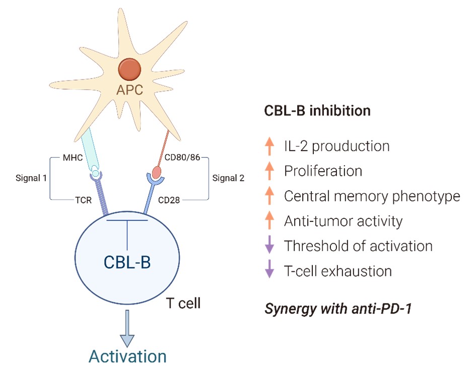
Currently, NX-1607 (also known as Cbl-b-IN-3), developed by Nurix Therapeutics, is the only Cbl-b inhibitor that has entered clinical research. Throughout this study, NX-1607 and Cbl-b-IN-3 will be used interchangeably to refer to the same compound. Our studies with Cbl-b-IN-3 have revealed its significant potential to enhance T and NK cell activation, underscoring the broader implications of Cbl-b inhibition in immuno-oncology therapies.
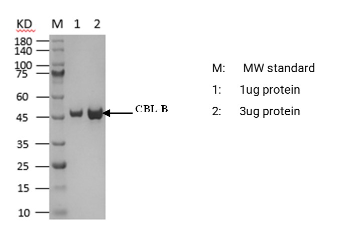
The Cbl-b protein was purified in-house at ICE Bioscience using an E. coli expression system. The purified protein demonstrated high purity and exhibited good activity (Figure 1).
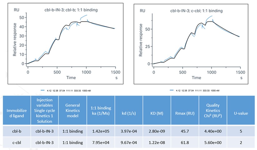
Small molecule inhibitors designed to target Cbl-b typically bind between the TKB domain and the LHR domain in its inactive conformation, stabilizing the protein in this non-active closed state. We used SPR analysis to evaluate the binding of Cbl-b-IN-3 to Cbl-b. As shown in Figure 2, Cbl-b-IN-3 bound with greater affinity to Cbl-b than to c-Cbl.
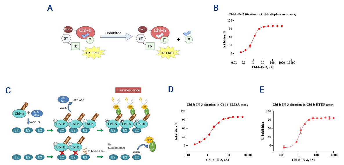
The displacement assay, HTRF assay, and ELISA all confirmed the dose-dependent inhibition of Cbl-b by Cbl-b-IN-3 in vitro (Figure 3).
The effect of Cbl-b-IN-3 on immune cells was measured using ICE Bioscience’s cell-based assays. We have also developed HiBiT cell lines for screening Cbl-b PROTAC molecules.
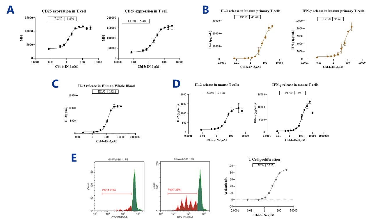
CD25 and CD69 are surface markers that are often used to assess T cell activation. In CD3-stimulated human T cells, inhibition of Cbl-b with Cbl-b-IN-3 promoted CD25 and CD69 expression (Figure 4A).
IL-2 and IFN-γ are crucial cytokines for T cell activation and the broader immune response. Inhibition of Cbl-b with Cbl-b-IN-3 promoted secretion of IL-2 and IFN-γ in both CD3 antibody-stimulated human T cells (Figure 4B) and mouse T cells (Figure 4D). Cbl-b-IN-3 also promoted the release of IL-2 in CD3 antibody-stimulated human whole blood (Figure 4C).
Anti-CD3/28 antibodies are often used in vitro to stimulate T cell activation and proliferation. Incubation of CellTrace™ Violet-labeled T cells with anti-CD3/28 antibodies and IL-2 in the presence of Cbl-b-IN-3 significantly enhanced T cell proliferation in a dose-dependent manner (Figure 4E).
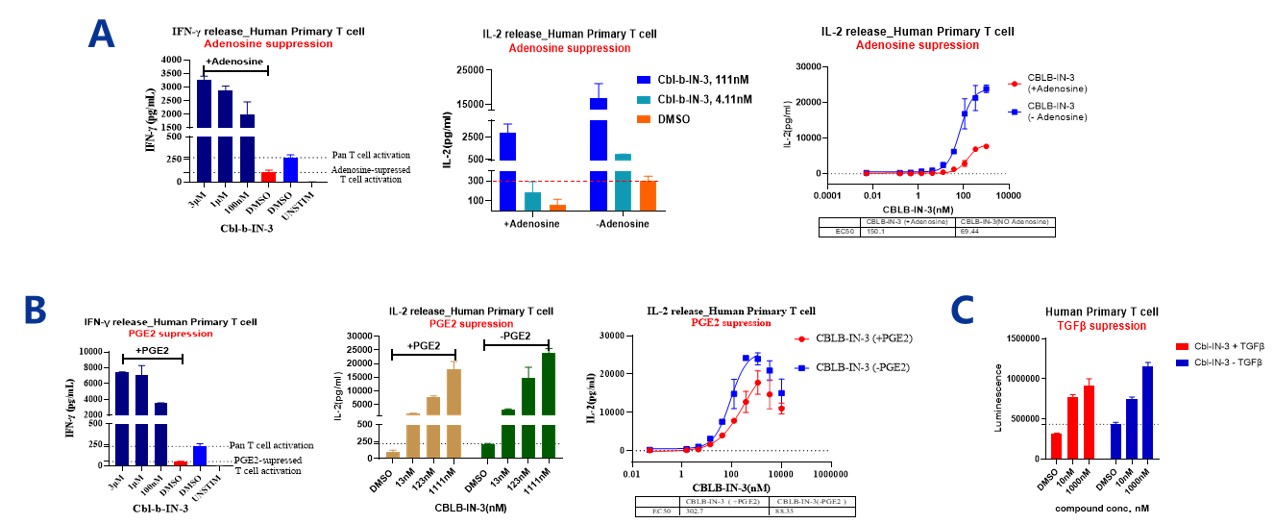
Several mechanisms within the tumor microenvironment (TME) can impair T cell responses that might otherwise control tumor growth. Notably, the accumulation of adenosine and prostaglandin E2 (PGE2) exerts a direct inhibitory effect on the early stages of T-cell activation, thereby facilitating tumor progression. Additionally, within the TME, TGF-β acts as a suppressor of T-cell proliferation, diminishes their effector functions, and hinders the maturation of T helper cells.
Adenosine and PGE2 suppressed T cell activation by inhibiting IL-2 and IFN-γ release in isolated T cells from PBMCs. However, a concurrent increase in the levels of IFN-γ and IL-2 cytokines was observed after treatment with Cbl-b-IN-3 combined with adenosine (Figure 5A) or PGE2 (Figure 5B). TGF-β induced suppression of human primary T cell proliferation, and Cbl-b-IN-3 reversed this effect by enhancing cell proliferation (Figure 5C).
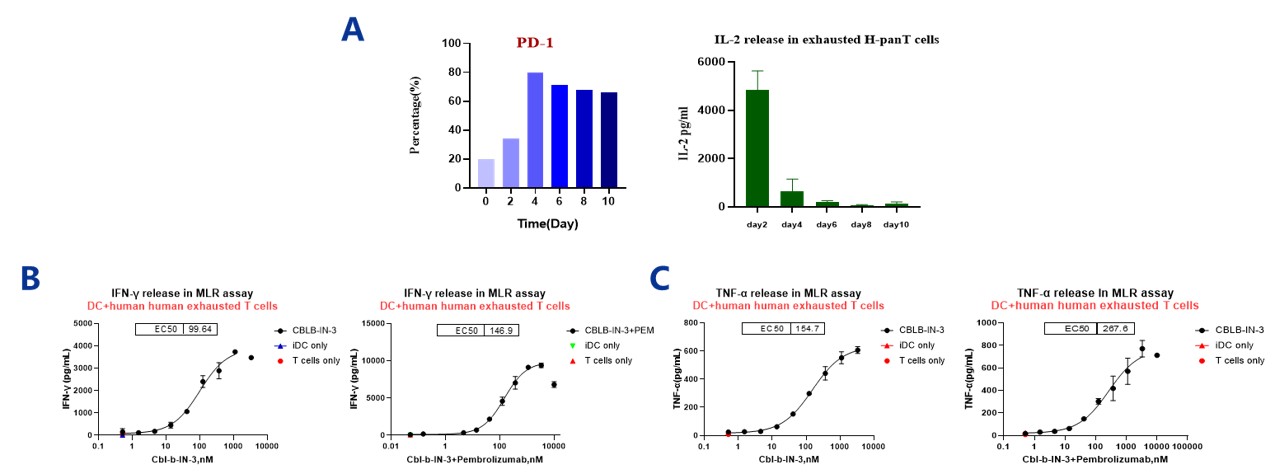
T-cell exhaustion can result from prolonged exposure of tumor cells to antigens, often occurring rapidly following tumorigenesis. This condition is typically marked by the high expression of inhibitory receptors such as CTLA-4, PD-1, TIM-3, LAG-3, and 2B4 on the T-cell surface. Additionally, it involves the loss of effector functions, including the production of cytokines like IFN-γ, IL-2, and TNF-α, and a reduction in the T-cells’ proliferative capacity. This state is characterized by a weakened T-cell response to antigens, rendering them less effective in providing T-cell help or eliminating target cells.
We induced exhausted T cells by stimulating them with an anti-CD3/CD28 activator. After 10 days, the increase in PD-1 expression and decrease in IL-2 release confirmed the induction (Figure 6A). The inhibition of Cbl-b with Cbl-b-IN-3 enhances T cell responses to dendritic cells (DCs), promoting the release of IFN-γ (Figure 6B) and TNF-α (Figure 6C) in mixed lymphocyte reaction (MLR) assays.
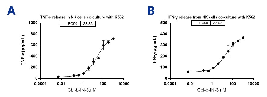
Tumors often develop resistance to the surveillance of the body’s innate immune cells, including natural killer (NK) cells. The E3 ubiquitin ligase Cbl-b has been recognized as a crucial regulator that restricts T cell activation and, more recently, NK cell activation. Consequently, NK cells deficient in Cbl-b exhibit an enhanced ability to combat tumor cells.
As shown in Figure 7, Cbl-b-IN-3 enhanced NK cell responses to target cells, leading to increased release of TNF-α and IFN-γ from NK cells when co-cultured with K562 cells.
Using our ready-to-use assays, we studied the effects of NX-1607 on T cells and NK cells under various conditions. The increase in CD25 and CD69 expression, along with the secretion of IL-2 and IFN-γ in CD3-stimulated human T cells, indicates the activation of T cells by NX-1607. NX-1607 can reverse the suppression of T cell activation exerted by adenosine, PGE2, and TGF-β. Additionally, NX-1607 can activate exhausted T cells in an MLR assay and enhance NK cell activation in a co-culture system. These results suggest a positive role for Cbl-b-IN-3 in cancer immunotherapy.
In addition to T cell and NK cell functional assays, scientists at ICE Bioscience have developed HiBiT cell lines to better screen Cbl-b PROTAC molecules by measuring their degradation effects. These HiBiT cell lines enable high-throughput screening in the early stages of drug discovery.
2026-08-20
2026-07-03
2026-05-22
2026-05-21
We value your inquiries and are here to provide you with tailored solutions for your drug discovery and development needs. Whether you have questions, require more information, or are interested in discussing potential collaborations, our team of experts is just a message away.
Feel free to reach out to us.

Address: Bldg 16, Yd 18, Kechuang 13th St, Etown, Tongzhou Dist, Beijing, 100176, China
Email: marketing@ice-biosci.com
Tel:+86-10-67809840


Get a quote

Go top