
EN CN


Neuroscience

Return

In Vivo Ischemic Stroke Models & Pharmacology Services

IN VIVO ISCHEMIC STROKE MODELS

Stroke is a leading cause of death and long-term disability worldwide, and the majority of stroke cases are ischemic in origin — triggered by the occlusion of a cerebral artery that abruptly cuts off oxygen and glucose supply to the brain. Despite decades of research, therapeutic options remain limited — thrombolysis and mechanical thrombectomy are only available within a narrow time window, and no clinically proven neuroprotective agent has yet reached routine practice. Validated in vivo models that faithfully recapitulate the temporal evolution of ischemic injury are the cornerstone of improving preclinical predictive value and de-risking clinical translation.

Leveraging deep expertise in neuroscience disease modeling, we have established a comprehensive in vivo ischemic stroke platform centered on middle cerebral artery occlusion (MCAO) in rats, offering two complementary focal cerebral ischemia models — transient MCAO (tMCAO) and permanent MCAO (pMCAO). Our models stably reproduce key ischemic phenotypes from acute hemodynamic disruption to chronic functional decline. We deliver comprehensive behavioral assessments including neurological severity score (mNSS), Rotarod test, Novel Object Recognition (NOR), and Morris Water Maze (MWM), complemented by TTC staining, HE staining, and immunofluorescence (IF) techniques. Combined with intraoperative Laser Doppler Flowmetry validation, these models support multi-layered efficacy evaluation and mechanistic dissection spanning hemodynamics, infarct evolution, neuroinflammatory cascades, and long-term neurorestoration.

MODEL SELECTION
Translational in vivo model selection for ischemic stroke drug discovery
Model TypeTypePrimary Pathological MechanismKey PhenotypesKey AdvantagesKey LimitationsOptimal Use Case
Transient MCAO (tMCAO)Focal cerebral ischemia with reperfusionTransient focal ischemia followed by reperfusion, resulting in ischemia–reperfusion injury, oxidative stress, BBB disruption, and neuroinflammationNeurological deficits, motor impairment, cerebral infarction, microglial activationClinically relevant model of reperfusion therapy; controlled reperfusion; ideal for acute neuroprotection studiesDoes not fully replicate spontaneous human reperfusion; infarct size may vary with surgical conditionsNeuroprotection, ischemia–reperfusion injury, BBB protection, anti-inflammatory therapies, reperfusion-targeted interventions
Permanent MCAO (pMCAO)Focal cerebral ischemia without reperfusionSustained focal ischemia without planned reperfusion; dominated by prolonged substrate deprivation, infarct-core expansion, collateral failure, delayed inflammation, and chronic remodelingPersistent neurological dysfunction, quantifiable chronic infarct, long-term motor and cognitive impairments, gliosisAvoids confounding reperfusion burst; useful for non-recanalized stroke and fixed-insult studies; direct surgical variants can confirm occlusion and yield reproducible lesions; allows chronic recovery and infarct-expansion analysisDoes not model recanalization, thrombectomy, or reperfusion-phase biology; direct occlusion may require craniectomy and advanced surgical skill; filament pMCAO may cause severe lesions and high disability; less representative of patients with spontaneous or therapeutic reperfusionStudying sustained occlusion, therapies that do not require restored flow, collateral dependence, infarct expansion, delayed inflammation, glial scarring, and chronic recovery
VALIDATED MODEL LIBRARY
Explore the model-specific validation workflow

Select a model below to review its biological rationale, experimental design, efficacy readouts, and representative data.

01Transient MCAO (tMCAO) Rat Model

The transient MCAO (tMCAO) rat model reproduces focal cerebral ischemia followed by reperfusion, closely mimicking ischemic stroke patients receiving recanalization therapy. The model induces reproducible neurological deficits, cerebral infarction, oxidative stress, and neuroinflammatory responses, making it a gold-standard platform for evaluating neuroprotective therapies against ischemia–reperfusion injury.

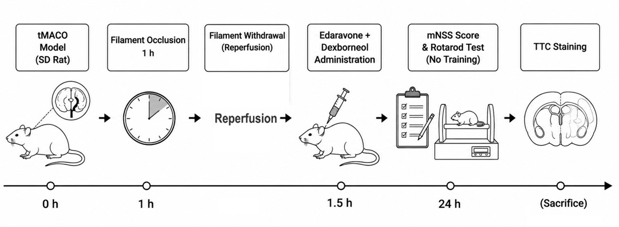
Experimental Flow Design

Experimental flow design of transient MCAO (tMCAO) rat model

Behavioral & Histological Characterization

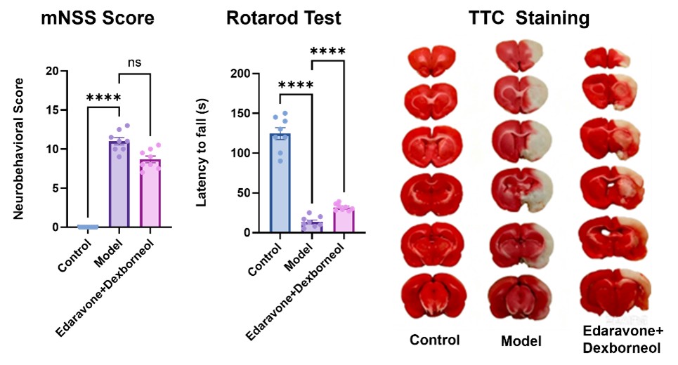
The tMCAO rat model successfully recapitulated key ischemic injury phenotypes, including neurological deficits in mNSS Score, impaired motor function in the Rotarod Test, and cerebral infarction in TTC staining. Edaravone and Dexborneol treatment significantly improved motor coordination and demonstrated trends toward improved neurological function and reduced infarct volume, supporting the utility of this model for evaluating neuroprotective therapies against ischemia–reperfusion injury.

Behavioral and histological characterization of tMCAO rat model

Pathological & Mechanistic Validation

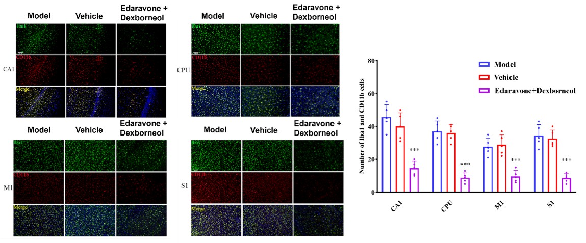
Iba1 & CD11b (Microglial activation): tMCAO induced marked microglial activation in the CA1, CPU, M1, and S1 regions, as evidenced by increased Iba1 and CD11b immunofluorescence. Edaravone and Dexborneol significantly reduced the expression of both markers across these brain regions, indicating effective attenuation of post-ischemic neuroinflammatory responses.

Iba1 and CD11b microglial activation staining in tMCAO model

02Permanent MCAO (pMCAO) Rat Model

Permanent middle cerebral artery occlusion (pMCAO) is a widely used focal cerebral ischemia model that mimics human ischemic stroke caused by persistent large vessel occlusion. The model induces permanent interruption of blood flow in the MCA territory, resulting in reproducible ischemic brain injury, infarction, neurological deficits, and long-term neuroinflammatory responses.

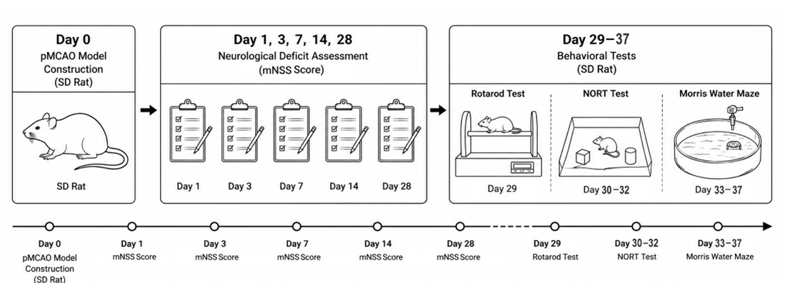
Experimental Flow Design

Experimental flow design of permanent MCAO (pMCAO) rat model

Intraoperative Hemodynamic Validation

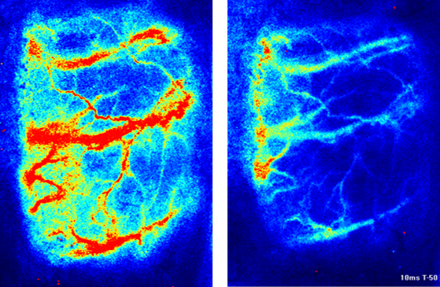
Laser Doppler Flowmetry: Successful induction of focal cerebral ischemia was intraoperatively confirmed by Laser Doppler Flowmetry, which demonstrated a marked and immediate reduction in regional cerebral blood flow (rCBF) within the MCA territory upon vessel occlusion. This hemodynamic validation serves as a critical quality control step to ensure consistent and reproducible model establishment across all study animals.

Laser Doppler Flowmetry validation of pMCAO rat model

Longitudinal Neurological & Structural Assessment

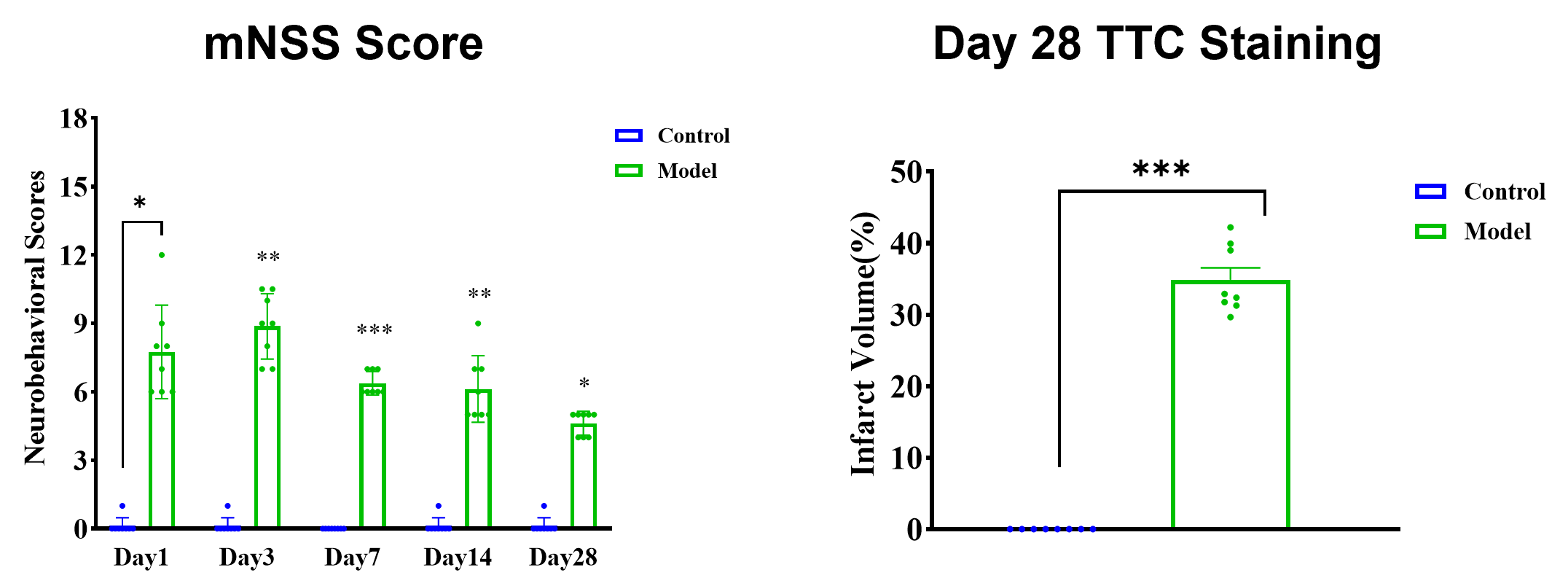
mNSS & TTC Staining: The pMCAO model produced sustained neurological impairment and persistent structural damage over the 28-day observation period. Longitudinal mNSS assessment revealed persistently elevated neurological severity scores in the Model group, indicating chronic neurological dysfunction without spontaneous recovery. Concordantly, TTC staining at Day 28 confirmed substantial and quantifiable cerebral infarct volume in the Model group, demonstrating that the permanent occlusion paradigm generates durable ischemic brain injury that remains structurally evident in the chronic phase.

Longitudinal mNSS scores and TTC staining of pMCAO rat model

Chronic Behavioral Characterization

Chronic functional consequences of permanent ischemia were comprehensively evaluated through a battery of behavioral tests spanning motor coordination (Rotarod test on Day 29), recognition memory (NOR test on Day 30–32), and spatial learning (MWM test on Day 33–37). The results demonstrate that pMCAO induces broad-spectrum, long-term behavioral impairments extending across both motor and cognitive domains, supporting its utility for evaluating neurorestorative and rehabilitative therapies.

Chronic behavioral characterization of pMCAO rat model

Histopathological Validation

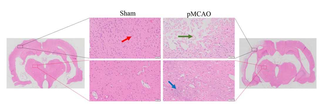
HE Staining: HE staining analysis confirms persistent ischemic brain injury after pMCAO. The sham group showed normal cortical structures with intact cells, abundant cytoplasm, and clear nucleoli (red arrows). In contrast, pMCAO rats at 28 days exhibited enlargement of the lateral ventricle, multifocal liquefactive necrosis, neuronal collapse and degeneration, fragmented and loosened tissue architecture (green arrows), and extensive inflammatory cell infiltration in the injured hemisphere (blue arrows), confirming sustained ischemic tissue damage and neuroinflammatory responses after pMCAO.

HE staining of ischemic brain injury in pMCAO rat model


Contact Us

We value your inquiries and are here to provide you with tailored solutions for your drug discovery and development needs. Whether you have questions, require more information, or are interested in discussing potential collaborations, our team of experts is just a message away.
Feel free to reach out to us.

We are a CRO service organization, not a hospital


Get a quote

Go top