
EN CN


Neuroscience

Return

In Vivo Parkinson's Disease Models & Pharmacology Services

In Vivo Parkinson's Disease Models
IN VIVO PARKINSON'S DISEASE MODELS

Parkinson's disease (PD) is the second most common neurodegenerative disorder, characterized by progressive loss of dopaminergic neurons in the substantia nigra pars compacta and a corresponding decline in striatal dopamine. The pathogenesis of PD is complex, involving mitochondrial dysfunction, oxidative stress, protein misfolding and aggregation, and neuroinflammation, with a markedly progressive disease course.

In vivo animal models are core tools for dissecting disease mechanisms, validating target druggability, and evaluating candidate drug efficacy. Models with distinct pathological mechanisms can be matched to different R&D stages, including symptomatic therapy, neuroprotection, and disease modification.

With extensive expertise in neurodegenerative disease modeling, we have established a comprehensive in vivo PD model platform covering multiple pathogenic mechanisms, including dopaminergic neurodegeneration, mitochondrial dysfunction, and α-Syn aggregation and propagation. We deliver comprehensive motor behavioral assessments, including Open Field Test (OFT), Grip Strength, and Rotarod test. Combined with pathological and molecular analyses, including ELISA and immunofluorescence (IF) staining, we offer comprehensive in vivo efficacy evaluation services, from model establishment and behavioral phenotyping to pathological and molecular mechanism analysis.

MODEL SELECTION
Translational in vivo model selection for Parkinson's disease drug discovery
Model TypePrimary MechanismKey AdvantagesKey LimitationsClinical RelevanceOptimal use case
6-OHDA Rat ModelOxidative stress (DAT/NET uptake)Robust, reproducible, unilateral design for within-subject controls; gold standard for cell transplantation studiesNo α-syn pathology; acute lesion; non-specific damage at injection siteSymptomatic therapy testing; cell transplantation; DBS developmentCell replacement therapy; symptomatic drug screening
MPTP Mouse ModelComplex I inhibition (DAT uptake)Well-characterized; clinically relevant mechanism (mitochondrial); scalable for drug screeningNo Lewy bodies; species limitationsMitochondrial drug screening; neuroprotection studiesMitochondrial-targeted neuroprotection; acute neurodegeneration mechanisms
PFF Injection Mouse ModelSeeded α-syn aggregationLewy body pathology; prion-like spread; Braak-like progression; most relevant for anti-α-syn therapiesExogenous protein; batch variability; compressed time course; injection inflammationAnti-aggregation therapy; immunotherapy; propagation studiesAnti-α-syn immunotherapy; prion-like propagation studies
AAV-α-Syn Mouse Modelα-Syn overexpression and aggregationProgressive pathology; dose-dependent severity; stage-specific phenotypes; validates α-syn lowering strategiesSupraphysiological overexpression; unilateral (typically); immune response to AAVDisease-modifying therapy; α-syn targeting; biomarker developmentProgressive neurodegeneration; stage-specific interventions; α-syn lowering therapies
VALIDATED MODEL LIBRARY
Explore the model-specific validation workflow

Select a model below to review its biological rationale, experimental design, efficacy readouts, and representative data.

016-OHDA-Induced Rat Model

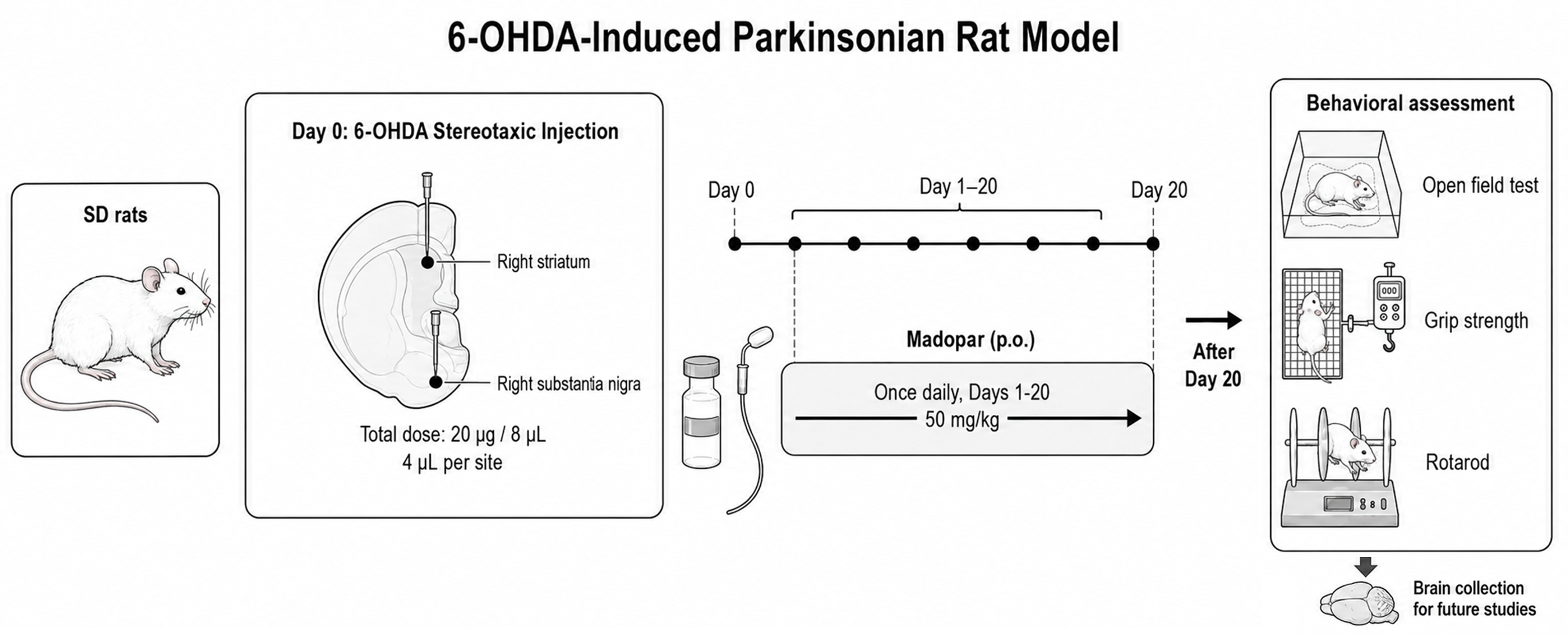
The unilateral 6-hydroxydopamine (6-OHDA) rat model is a well-established toxin-based model of Parkinson's disease. Stereotaxic injection of 6-OHDA into the right striatum and substantia nigra induces selective degeneration of the nigrostriatal dopaminergic pathway, resulting in motor impairment, reduced tyrosine hydroxylase (TH) expression, and neuroinflammatory activation. Following model establishment, Madopar is orally administered once daily for 20 consecutive days, followed by comprehensive behavioral testing (OFT, Grip Strength, Rotarod) and brain histopathological analysis.

Experimental flow design

Experimental Flow Design of 6-OHDA-Induced Rat Model

Behavioral characterization

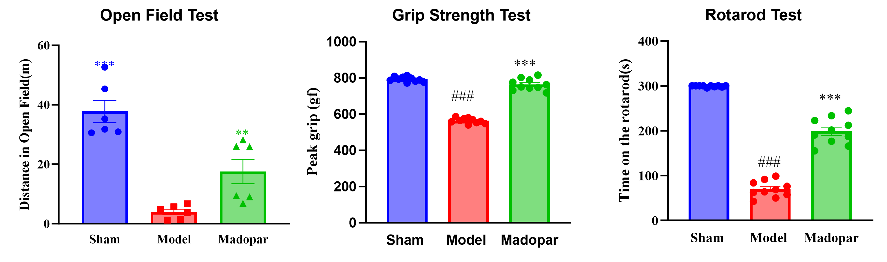
The 6-OHDA-induced Parkinson's disease rat model successfully recapitulated key motor deficits, including reduced locomotor activity, decreased grip strength, and impaired motor coordination. These abnormalities were effectively ameliorated by Madopar, confirming the model's responsiveness to dopaminergic replacement therapy.

Behavioral characterization of the 6-OHDA-induced rat model

Pathological & IF analysis

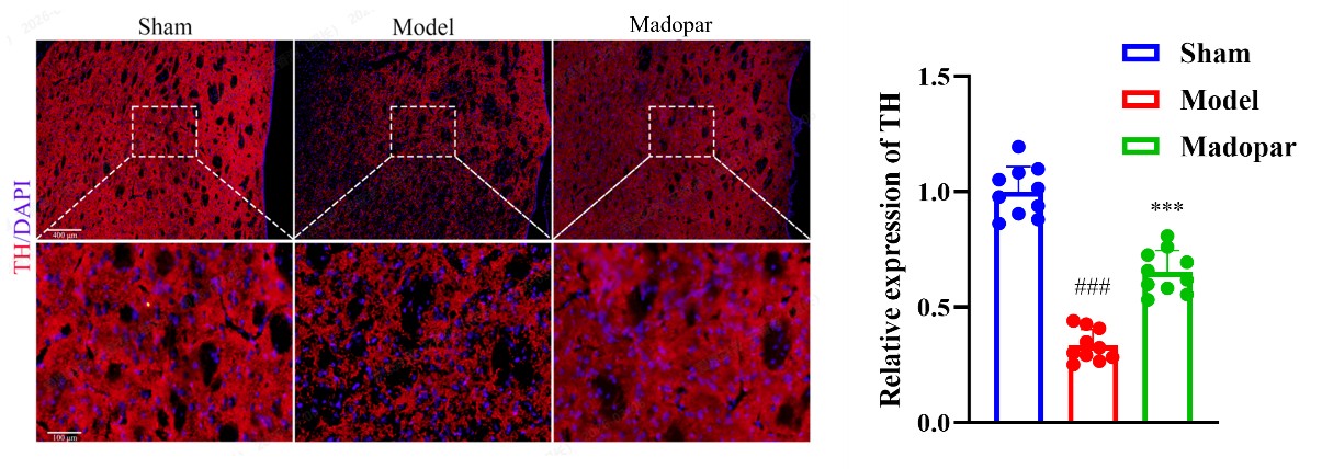
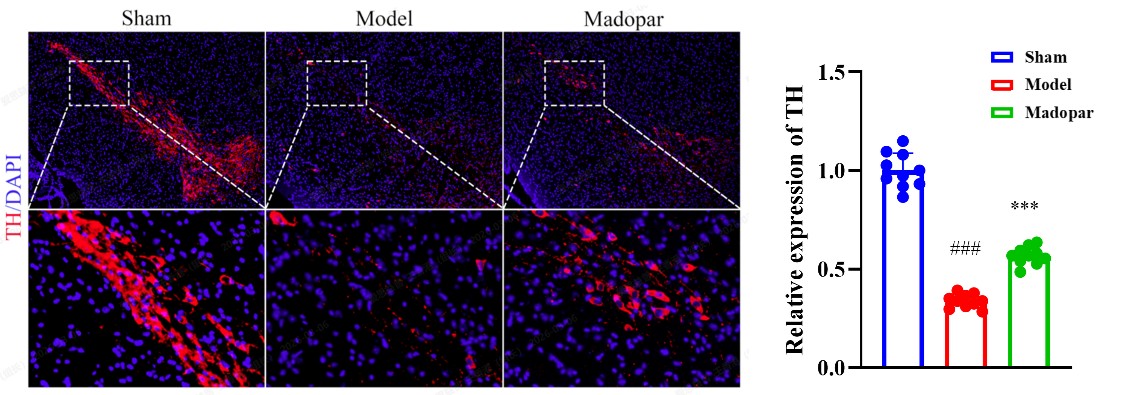
TH IF in the Striatum: Reduced TH signal in the model group, indicating dopaminergic terminal damage; TH expression was partially restored following oral Madopar treatment.

TH IF in the Striatum

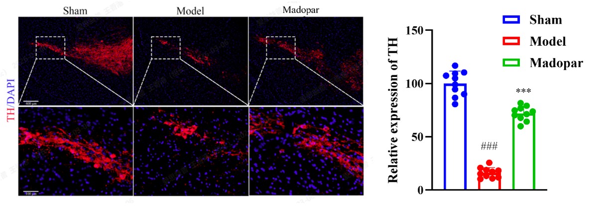
TH IF in the Substantia Nigra: Loss of dopaminergic neurons in the model group; increased TH signal with Madopar, indicating partial neuroprotection.

TH IF in the Substantia Nigra

Neuroinflammation evaluation

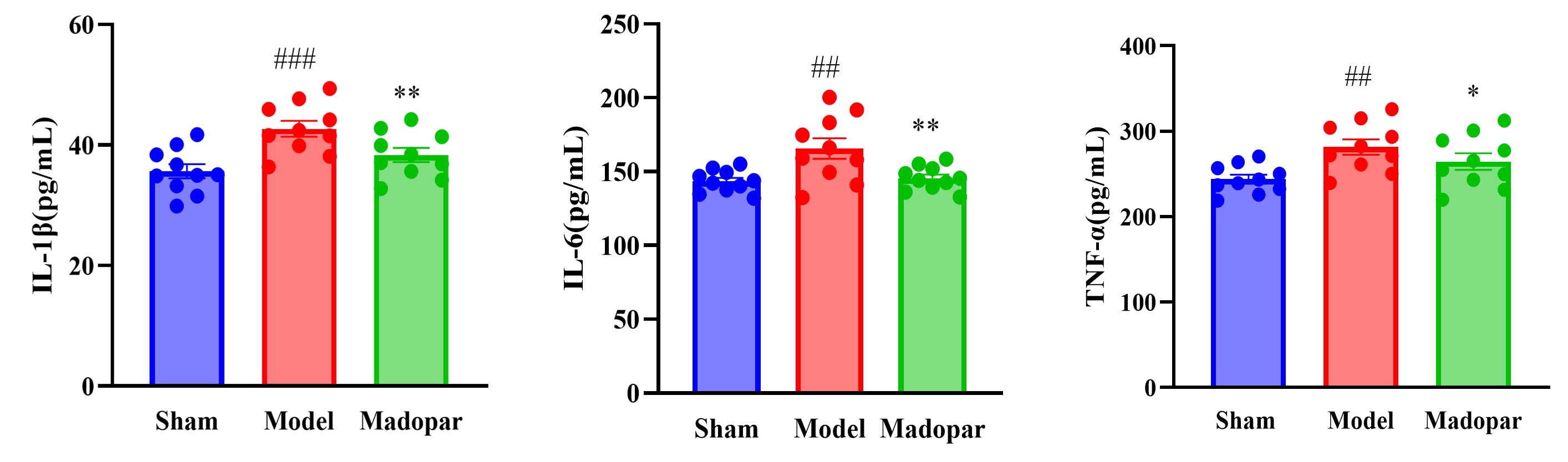
Inflammatory factors in CSF: 6-OHDA induced significant elevation of inflammatory factors (IL-1β, IL-6, TNF-α) in cerebrospinal fluid (CSF) by ELISA; oral Madopar attenuated this inflammatory response.

Inflammatory factors in CSF

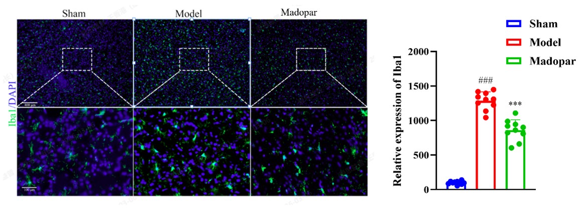
Iba1 IF in the Striatum: Increased Iba1 signals in the model group indicated microglial activation; Madopar reduced microglial activation in the striatum.

Iba1 IF in the Striatum

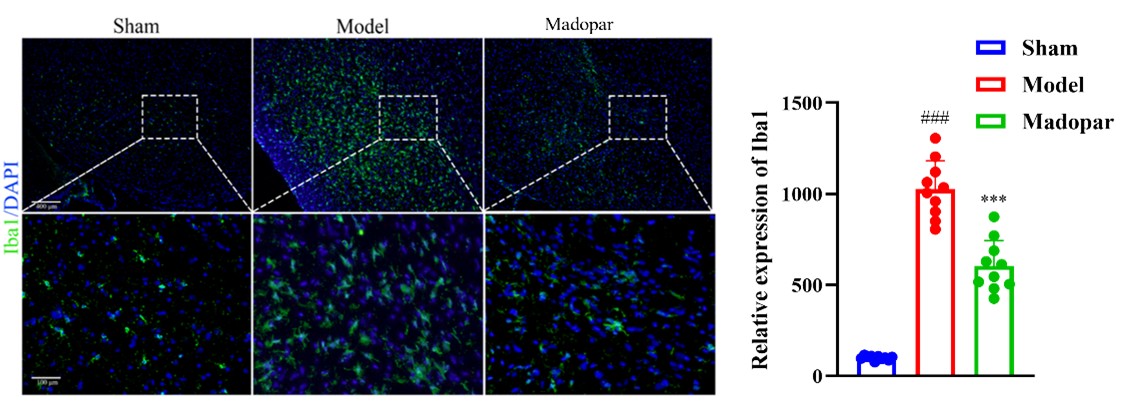
Iba1 IF in the Substantia Nigra: Consistent microglial activation in the model group observed in the substantia nigra; Madopar similarly attenuated microglial changes.

Iba1 IF in the Substantia Nigra

02Subacute MPTP-Induced Mouse Model

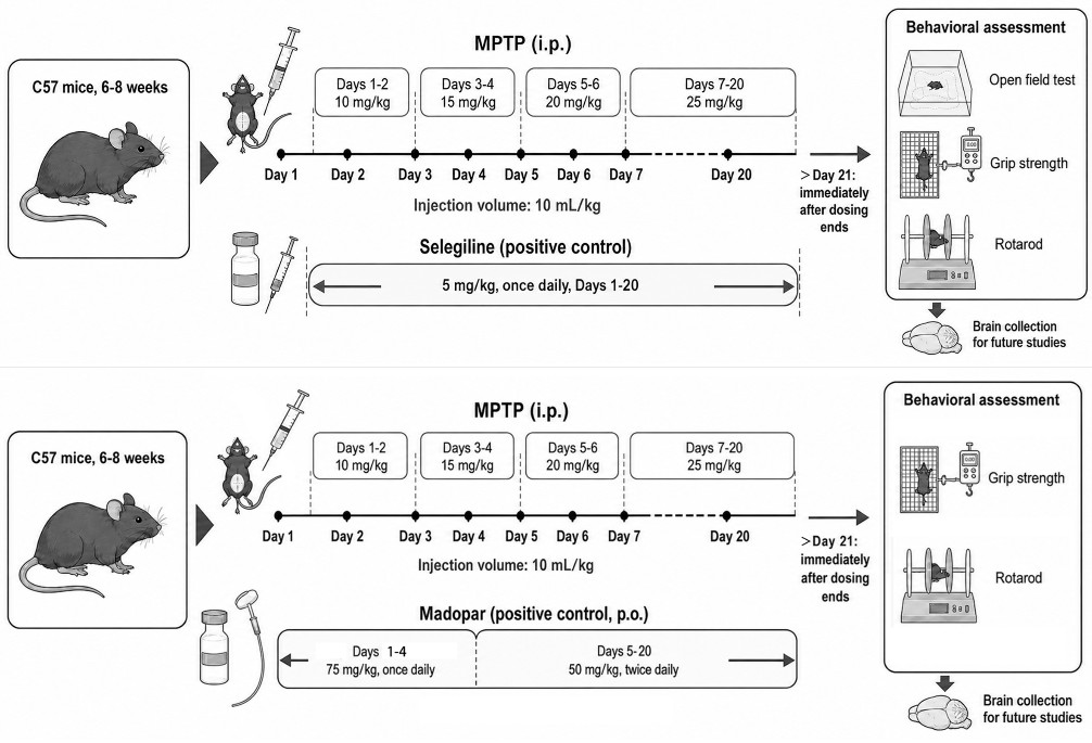
Repeated escalating intraperitoneal administration of MPTP in C57BL/6 mice induces progressive nigrostriatal dopaminergic dysfunction. MPTP is converted to MPP⁺, which preferentially enters dopaminergic neurons and impairs mitochondrial complex I, leading to oxidative stress and dopaminergic neurodegeneration. To validate assay sensitivity, two mechanistically distinct reference compounds were incorporated: Selegiline and Madopar. Following model establishment and reference compound treatment, animals underwent behavioral testing (OFT, Grip Strength, and Rotarod) and subsequent brain histopathological analysis.

Experimental flow design

Experimental Flow Design of MPTP-Induced Mouse Model

Pharmacological validation with Selegiline

Behavioral characterization

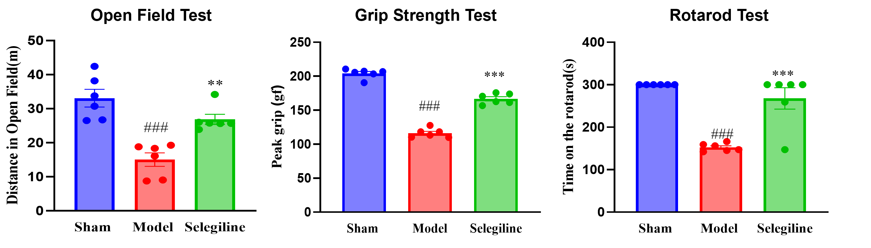
Repeated MPTP administration induced clear motor impairment, including reduced locomotor activity, decreased grip strength, and impaired motor coordination. Selegiline significantly attenuated these behavioral deficits; however, performance was not fully restored to control levels.

Behavioral characterization with Selegiline treatment

Pathological & IF analysis

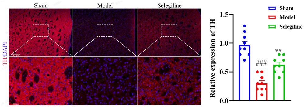
TH IF in the Striatum: Reduced TH expression in the model group, indicating dopaminergic terminal damage; Selegiline partially preserved TH signals.

TH IF in the Striatum after Selegiline treatment

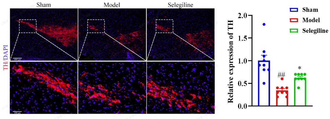
TH IF in the Substantia Nigra: Loss of dopaminergic neurons in the model group; TH signals were significantly but incompletely restored following Selegiline treatment.

TH IF in the Substantia Nigra after Selegiline treatment

Pharmacological validation with Madopar

Behavioral characterization

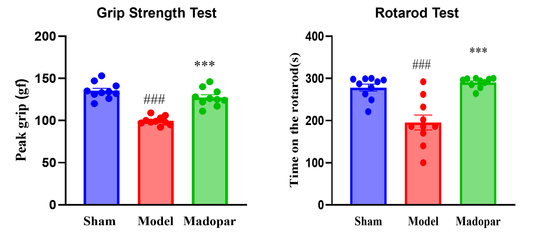
Subacute MPTP treatment induced significant motor deficits, including reduced grip strength and impaired rotarod performance; these abnormalities were effectively ameliorated by Madopar.

Behavioral characterization with Madopar treatment

Pathological & IF analysis

TH IF in the Substantia Nigra: MPTP-associated TH loss was observed in the model group; Madopar significantly ameliorated TH loss in the substantia nigra, supporting the responsiveness of this model to dopaminergic replacement therapy.

TH IF in the Substantia Nigra after Madopar treatment

03AAV9-CMV-α-Synuclein-Induced Mouse Model

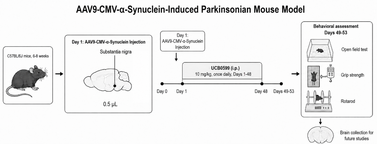
Stereotaxic delivery of AAV9-CMV-α-synuclein into the substantia nigra induces sustained α-synuclein overexpression and progressive nigrostriatal dopaminergic dysfunction. This model captures key features of synucleinopathy, including motor impairment, dopaminergic neuronal injury, and pathogenic α-synuclein accumulation. Following model establishment, UCB0599 is intraperitoneally (i.p.) administered once daily for 48 consecutive days, followed by comprehensive behavioral testing (OFT, Grip Strength, Rotarod) and brain histopathological analysis.

Experimental flow design

Experimental Flow Design of AAV9-CMV-α-Synuclein-Induced Mouse Model

Behavioral characterization

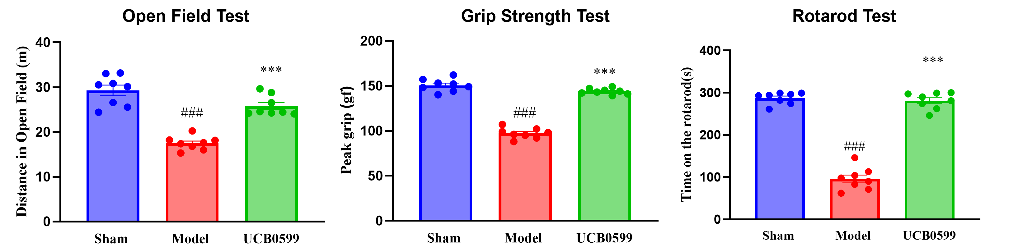
The AAV9-CMV-α-synuclein-induced Parkinson's disease mouse model successfully recapitulated key motor deficits, including reduced locomotor activity, decreased grip strength, and impaired motor coordination. These abnormalities were effectively ameliorated by UCB0599, confirming the model's responsiveness to α-synuclein-targeted therapy.

Behavioral characterization of the AAV-α-Syn mouse model

Pathological & IF analysis

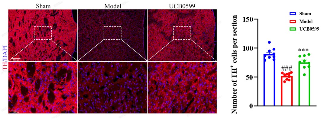
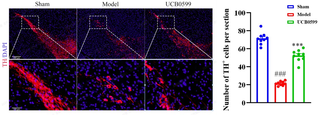
TH IF in the Striatum: Reduced TH signal in the model group, indicating dopaminergic terminal damage; TH fluorescence was increased following UCB0599 treatment.

TH IF in the Striatum after UCB0599 treatment

TH IF in the Substantia Nigra: Loss of dopaminergic neurons in the model group; TH signal was restored with UCB0599, indicating neuroprotection.

TH IF in the Substantia Nigra after UCB0599 treatment

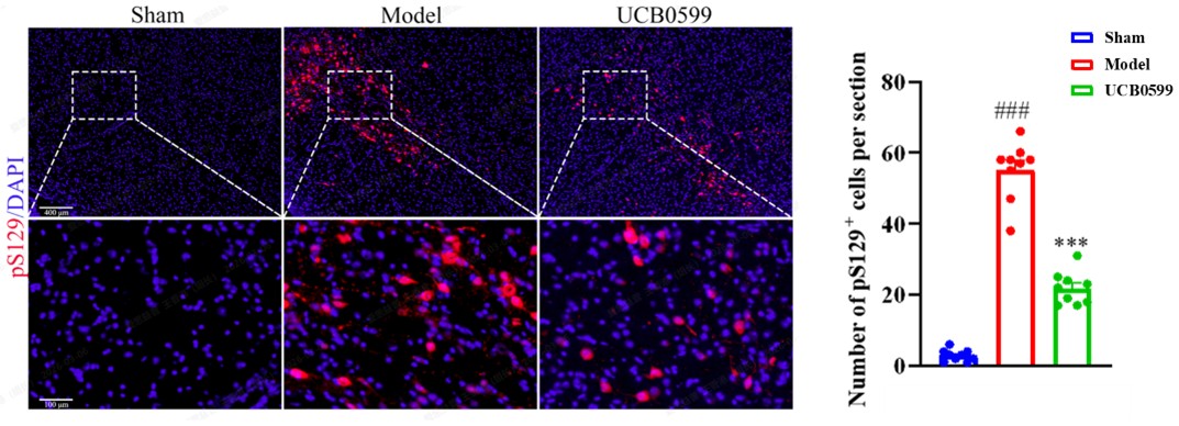
pSer129 IF in the Substantia Nigra: Marked accumulation of pathological pSer129-α-synuclein in the model group; UCB0599 significantly reduced pSer129-α-synuclein signals, supporting its potential to attenuate α-synuclein-associated nigrostriatal pathology.

pSer129 IF in the Substantia Nigra after UCB0599 treatment

04PFF-Induced Parkinsonian Mouse Model

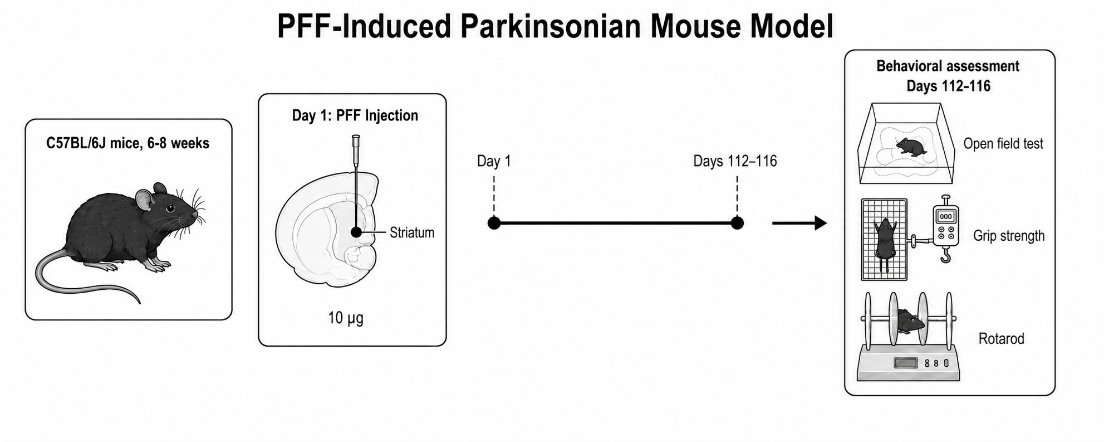
Intrastriatal injection of α-synuclein preformed fibrils (PFFs) triggers progressive α-synuclein pathology and nigrostriatal dysfunction, recapitulating the prion-like seeding and spreading of pathological α-syn observed in Parkinson's disease. Motor performance was evaluated 112 days after PFF administration using Open Field, Grip Strength, and Rotarod tests — a timepoint capturing the late-onset, progressive nature of PFF-induced pathology.

Experimental flow design

Experimental Flow Design of PFF-Induced Mouse Model

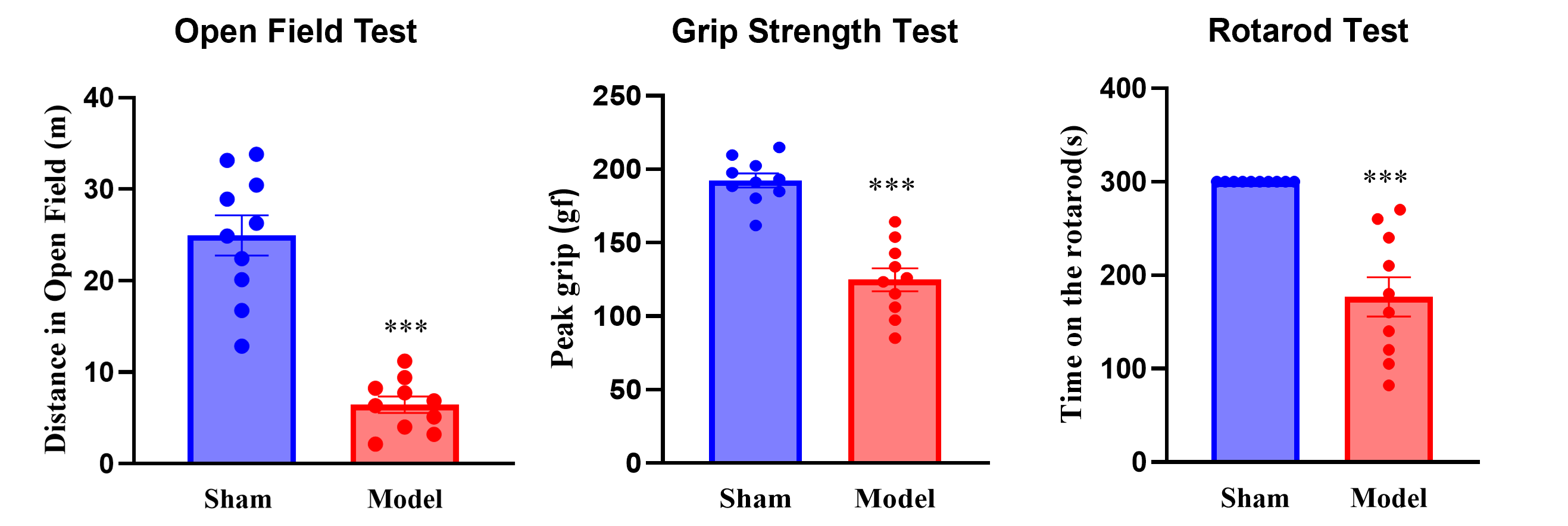
Behavioral characterization

The PFF-induced mouse model showed impaired locomotor activity, muscle strength, and motor coordination, demonstrating robust late-onset motor deficits in this progressive synucleinopathy model.

Behavioral characterization of the PFF-induced mouse model


Contact Us

We value your inquiries and are here to provide you with tailored solutions for your drug discovery and development needs. Whether you have questions, require more information, or are interested in discussing potential collaborations, our team of experts is just a message away.
Feel free to reach out to us.

We are a CRO service organization, not a hospital


Get a quote

Go top