IN VITRO ALZHEIMER'S DISEASE MODELS
Alzheimer's disease (AD) is the most prevalent neurodegenerative disorder worldwide. Its pathogenesis involves a cascade of interconnected mechanisms, including β-amyloid (Aβ) deposition, tau hyperphosphorylation, glutamatergic excitotoxicity, oxidative stress damage, and neuroinflammation — factors that have long created major barriers to efficient, translatable drug discovery.
In vitro cell models serve as a cornerstone of early-stage AD drug development. They enable precise recapitulation of key AD pathological phenotypes at the cellular level, providing a rapid, high-throughput platform for efficacy screening and mechanism-of-action (MoA) validation of candidate compounds, effectively shortening R&D cycles and reducing the cost of in vivo studies.
We offer well-validated in vitro AD models, covering the major pathological mechanisms to meet diverse R&D scenarios. All models are systematically validated via standardized CCK-8 cell viability assay and immunofluorescence staining, ensuring robust, reproducible and biologically relevant data for AD drug discovery.
VALIDATED MODEL LIBRARY
Explore the model-specific validation workflow
Select a model below to review its biological rationale, experimental design, efficacy readouts, and representative data.
01Aβ1-42-Induced SH-SY5Y Cell Model
The Aβ1-42-induced SH-SY5Y cell model recapitulates amyloid toxicity — a critical pathogenic event driving AD progression. Through concentration gradient screening, 20 μM Aβ1-42 was identified as the optimal modeling concentration, stably inducing neuronal injury and providing a robust platform for evaluating neuroprotective compounds against amyloid toxicity.
Model Optimization
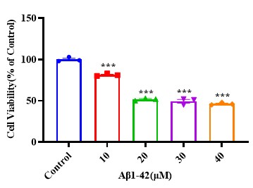
Concentration-gradient validation was performed via the CCK-8 cell viability assay. 20 μM Aβ1-42 was selected as the optimal induction concentration, supporting consistent and robust induction of amyloid-mediated neuronal injury.
Pharmacological Validation
Three reference compounds with distinct mechanisms were selected for efficacy validation:
✅ MCC950: A selective NLRP3 inflammasome inhibitor that suppresses neuroinflammation
✅ 2-AG (2-Arachidonoylglycerol): An endogenous endocannabinoid that activates CB1/CB2 receptors to exert neuroprotective and anti-inflammatory effects
✅ 7-BIO: A GSK-3 inhibitor that modulates tau phosphorylation and neuronal survival signaling
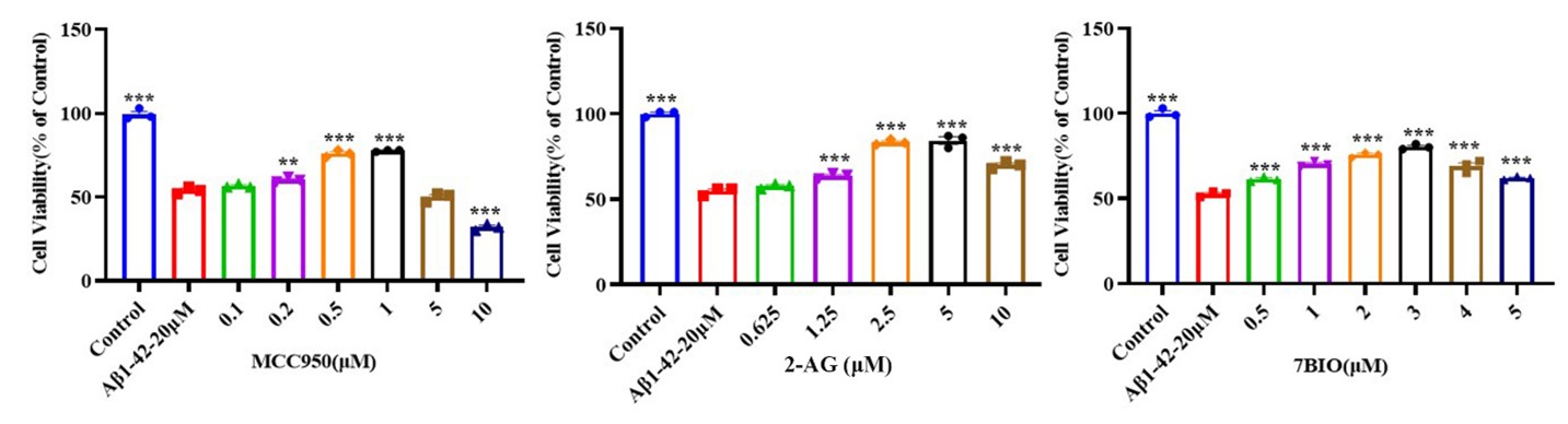
The results demonstrated that all three compounds exerted concentration-dependent neuroprotective effects and effectively ameliorated Aβ1-42-induced cellular injury, supporting the identification of optimal dosing conditions in AD drug discovery.
Mechanistic Validation
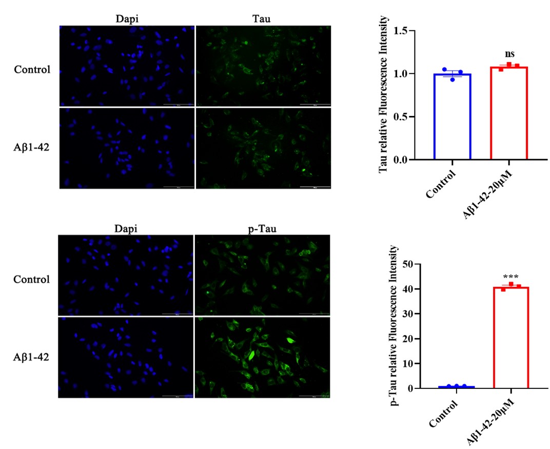
Immunofluorescence staining revealed that total Tau expression remained unchanged following Aβ1-42 treatment. However, Aβ1-42 significantly increased phosphorylated Tau (P-Tau) expression, indicating the induction of AD-related tau pathology without altering total Tau abundance.
02Glutamate-Induced SH-SY5Y Cell Model
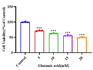
The glutamate-induced SH-SY5Y cell model recapitulates glutamatergic excitotoxicity — a critical pathogenic event driving AD progression. Concentration gradient screening was carried out via the CCK-8 cell viability assay, and 20 mM glutamate was selected as the optimal induction concentration for pharmacological evaluation.
Model Optimization
This model recapitulates glutamatergic excitotoxicity. Concentration gradient screening was carried out via the CCK-8 cell viability assay, and 20 mM glutamate was selected as the optimal induction concentration, supporting consistent and robust induction of excitotoxic neuronal injury.
Pharmacological Validation
Three reference compounds with distinct mechanisms of action were tested to validate the model's pharmacological responsiveness, with efficacy measured by the CCK-8 cell viability assay:
✅ 7-BIO: A GSK-3 inhibitor that suppresses tau hyperphosphorylation and promotes neuronal survival
✅ Donepezil: An acetylcholinesterase (AChE) inhibitor that enhances cholinergic neurotransmission, an approved symptomatic treatment for AD
✅ Resveratrol: A natural polyphenolic antioxidant that alleviates oxidative stress, inhibits neuroinflammation and activates SIRT1-mediated neuroprotective pathways
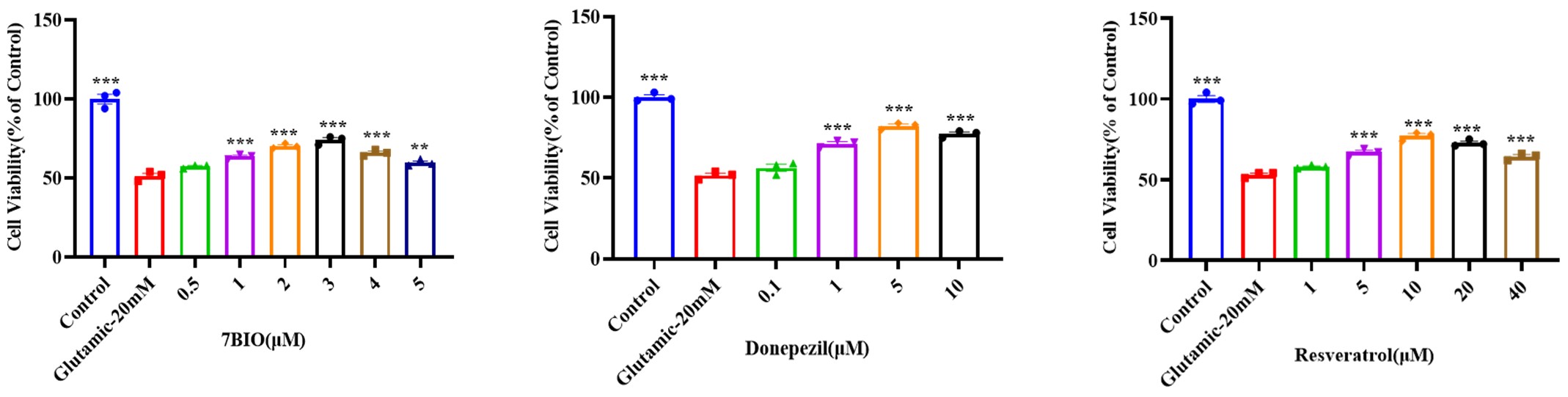
The results showed that all three compounds exhibited significant neuroprotective effects against glutamate-induced excitotoxicity, verifying the model's capability to detect efficacy across compounds with diverse modes of action.
Mechanistic Validation
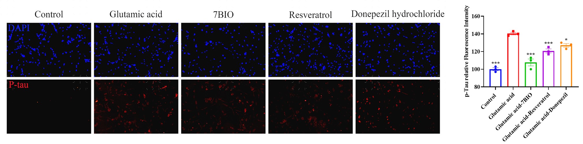
Immunofluorescence staining was applied to verify modulation of tau pathology. Glutamate stimulation markedly upregulated intracellular P-Tau expression, while 7-BIO, donepezil and resveratrol all significantly attenuated glutamate-induced tau hyperphosphorylation. These findings confirm the biological relevance of the model and support its use in mechanism-of-action dissection of candidate compounds.
03Primary Hippocampal Neuron AD Model
To complement cell line models with a more physiologically relevant system, we established a panel of three AD injury models using primary hippocampal neurons, induced by Aβ1-42, okadaic acid (OA) and glutamate respectively. All models are systematically validated via standardized CCK-8 cell viability assay and immunofluorescence staining, ensuring robust, reproducible and biologically relevant data for AD drug discovery.
Model Optimization
Concentration-gradient validation was performed via the CCK-8 cell viability assay. All three models exhibited concentration-dependent neuronal injury, with reduced cell viability. Following systematic optimization, the optimal inducing concentrations were determined as 20 μM for Aβ1-42, 1 nM for okadaic acid, and 20 mM for glutamate.
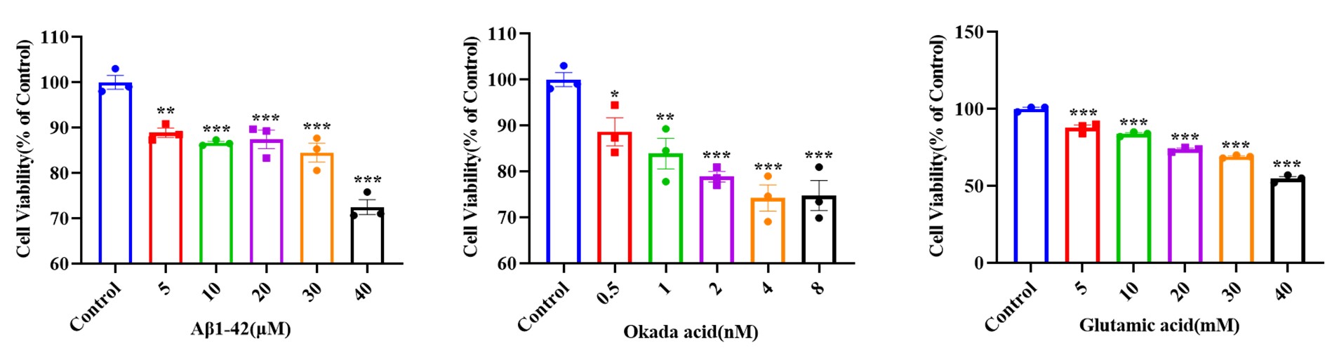
Phenotypic Characterization
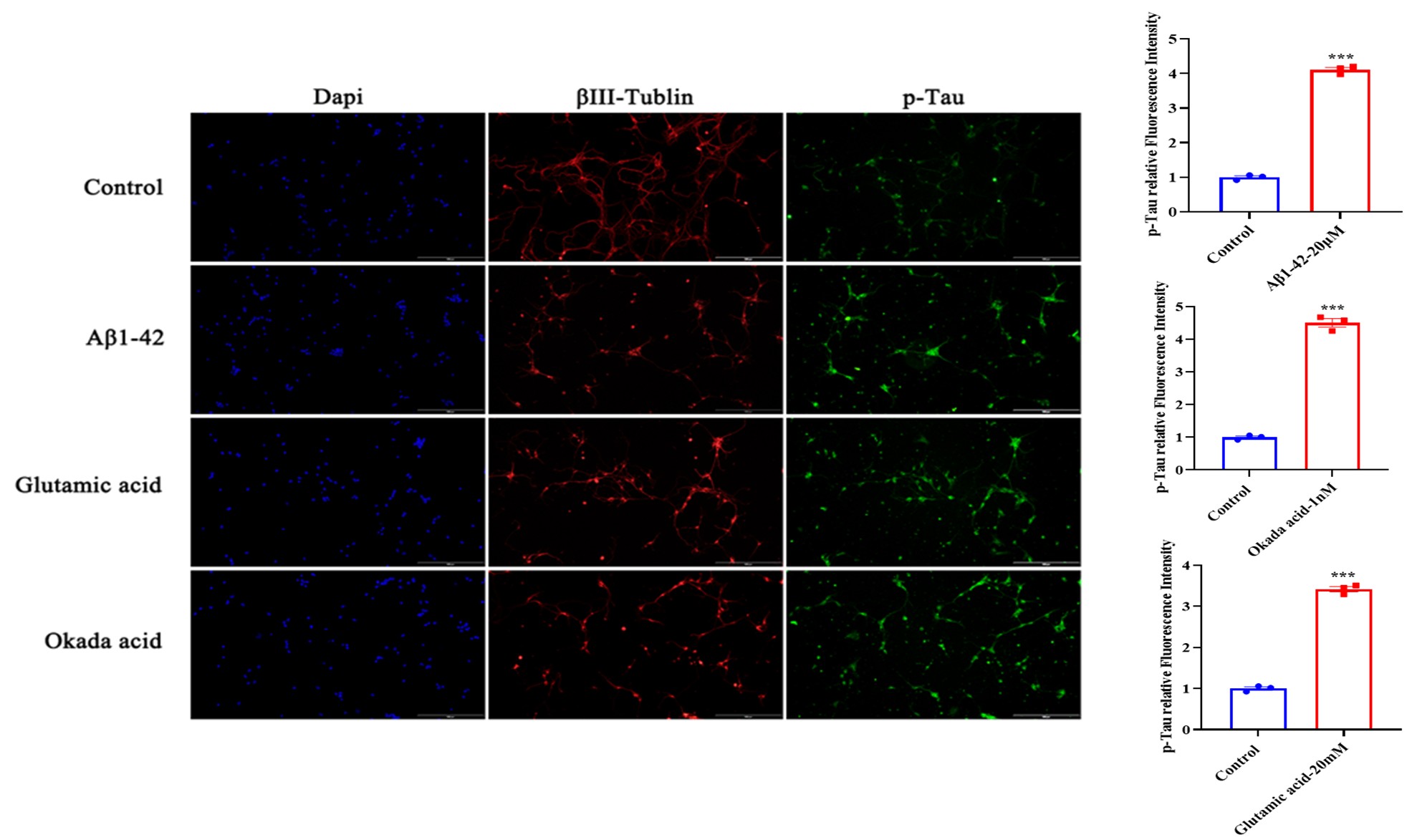
Immunofluorescence staining was utilized to profile phenotypic and pathological alterations, including downregulated expression of the neuronal marker βIII-tubulin and elevated tau phosphorylation levels, successfully recapitulating core neurodegenerative phenotypes of AD.