
EN CN


Neuroscience

Return

In Vitro Stroke Models & Assay Services

IN VITRO STROKE MODELS

Stroke remains one of the leading causes of death and long-term disability worldwide, driven by complex pathophysiological cascades including ischemic energy failure, excitotoxicity, oxidative stress, neuroinflammation, and programmed cell death. Robust in vitro models that faithfully recapitulate key stroke-related pathologies are essential for accelerating mechanistic understanding and therapeutic discovery.

Our platform employs a panel of well-characterized cellular models that target distinct yet interconnected mechanisms of ischemic brain injury. From oxygen-glucose deprivation (OGD) simulating ischemia/reperfusion, to glutamate-induced excitotoxicity modeling neuronal overstimulation, and LPS-activated microglial inflammation reflecting secondary immune responses — each model is paired with multi-parameter phenotypic and molecular readouts to deliver mechanistic insights and pharmacological validation.Our assay portfolio includes CCK-8 cell viability assay, LDH cytotoxicity assay, cellular ROS detection, Griess assay for nitric oxide quantification, immunofluorescence staining, ELISA for cytokine profiling, and neuronal morphological analysis — enabling comprehensive evaluation of compound efficacy across cell survival, oxidative stress, inflammatory response, apoptosis, and neurite integrity.

These models serve as powerful, scalable tools for compound screening, mechanism-of-action (MoA) studies, and neuroprotective candidate validation prior to in vivo translation.

MODEL SELECTION
Mechanism-driven in vitro model selection for stroke drug discovery
Modeling MethodPrimary pathological mechanism modeledCommonly used cell typeKey molecular pathwaysMain phenotypic readoutOptimal use case
OGD-inducedIschemic substrate deprivation; energy failure, oxidative stress, mitochondrial dysfunction, ER stress, inflammasome activation, apoptosis or parthanatosSH-SY5Y neuroblastoma cells, primary hippocampus neuronATP depletion, ROS, mitochondrial depolarization, ER stress ATF4/ATF6/PERK/IRE1, PARP-1/PAR/AIF, NF-κB, NLRP3, Nrf2/HO-1, AMPKMTT/CCK-8, LDH, trypan blue, ROS, MDA/GSH, JC-1 mitochondrial potentialScreening compounds against ischemia-like neuronal energy failure; testing mitochondrial, ER-stress, parthanatos, inflammasome, and antioxidant mechanisms
Glutamate-inducedExcitotoxic and oxidative neuronal injury without necessarily reproducing full ischemic deprivationSH-SY5Y cells; primary cortical or hippocampal neuronsROS, lipid and protein oxidation, mitochondrial dysfunction, Bax/Bcl-2, cytochrome c, caspase-3, JNK and p38 MAPK; receptor dependence must be validated in the chosen clone and differentiation stateViability, LDH, ROS, MDA, protein carbonyls, SOD/catalase, mitochondrial membrane potential, Annexin V/PI, Bax/Bcl-2/cleaved caspase-3Isolating excitotoxic/oxidative injury and testing mitochondrial or MAPK-targeted neuroprotection; not sufficient alone to infer protection from full ischemia
LPS-inducedMicroglial inflammatory amplification and innate immune activation; models secondary neuroinflammation rather than ischemic energy failureMurine BV2 microgliaTLR4/MyD88/IRAK, IκBα/NF-κB, ERK/JNK/p38 MAPK, iNOS/NO, COX-2/PGE2, IL-1β, TNF-α, IL-6, miR-155, STAT3 and NLRP3 depending on protocolGriess NO, PGE2, TNF-α, IL-1β, IL-6, microglial morphology, neuronal death in cocultureTesting anti-inflammatory mechanisms, microglial signaling, and immune-mediated neuronal injury; should be paired with OGD or in vivo models for stroke relevance
VALIDATED MODEL LIBRARY
Explore the model-specific validation workflow

Select a model below to review its biological rationale, experimental design, efficacy readouts, and representative data.

01OGD/R-Induced SH-SY5Y Cell Model

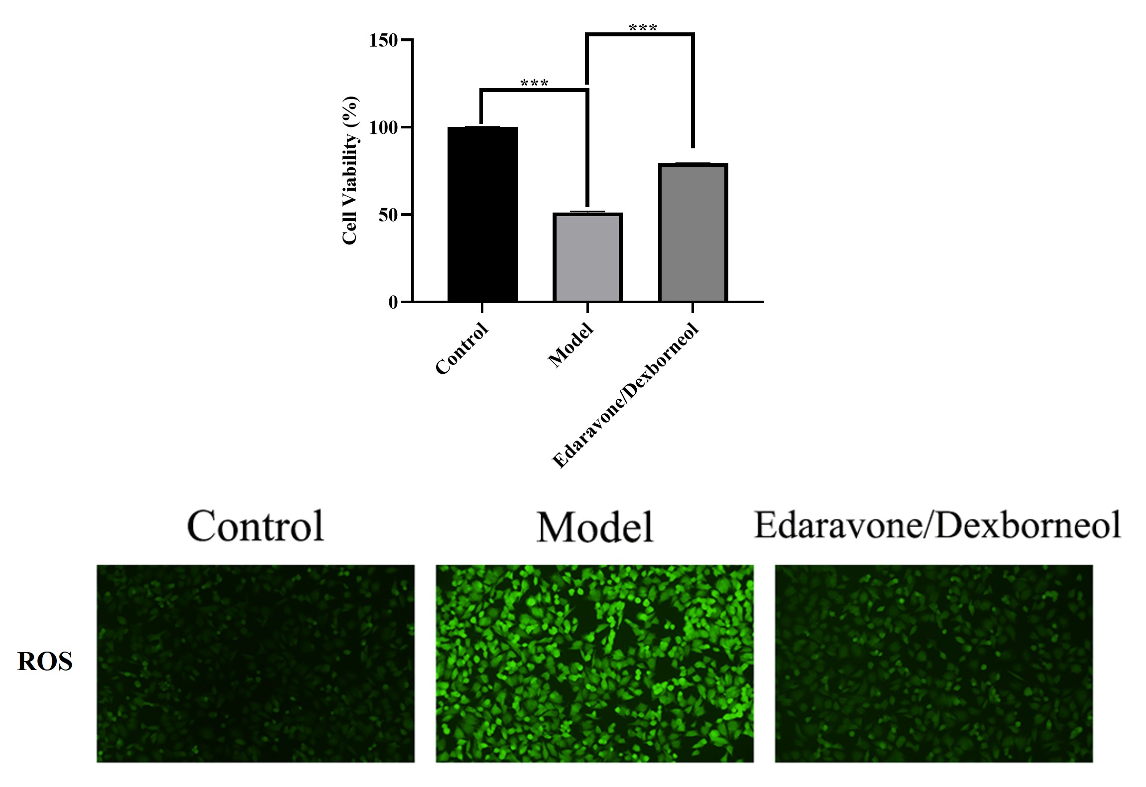
In the oxygen-glucose deprivation/reoxygenation (OGD/R) mode, we offer CCK-8 cell viability assay and cellular ROS detection to evaluate neuroprotective effects of test compounds. The OGD/R model successfully reproduced ischemia/reperfusion-induced neuronal injury, characterized by reduced cell viability and excessive oxidative stress. The Edaravone/Dexborneol treatment effectively improved cell survival and attenuated oxidative stress.

CCK-8 cell viability and cellular ROS detection in OGD/R model

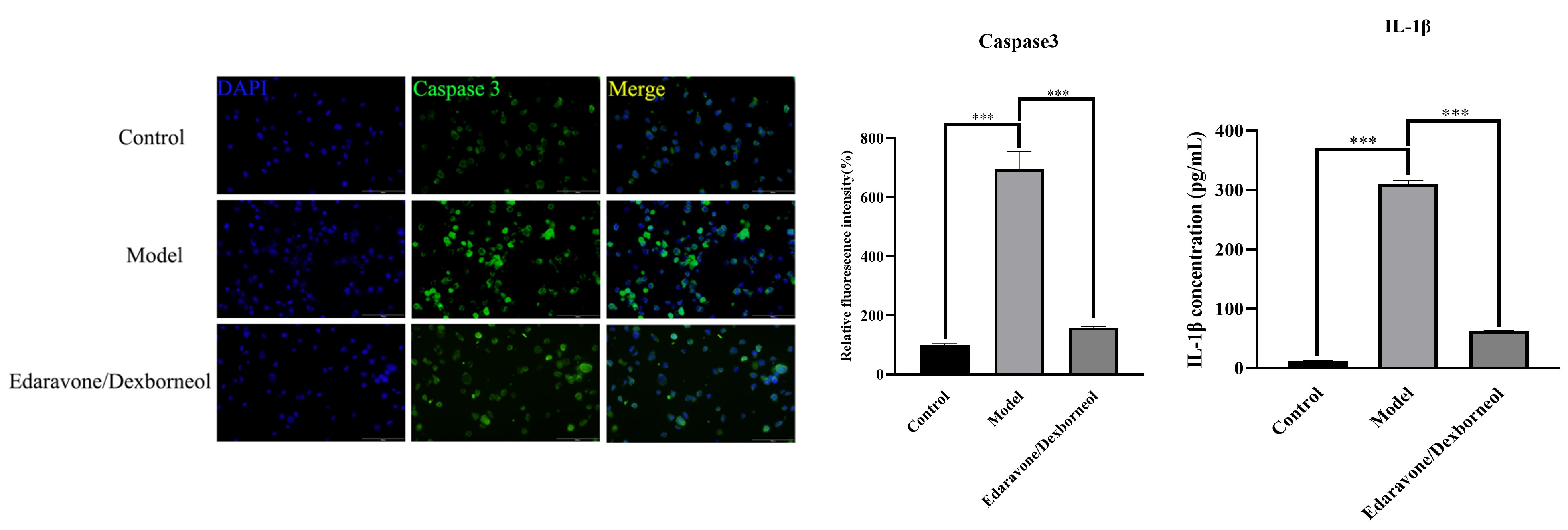
Mechanistically, OGD/R activated apoptosis-associated signaling by Caspase-3 immunofluorescence and inflammatory responses by IL-1β ELISA, whereas Edaravone/Dexborneol effectively suppressed both neuronal apoptosis and the inflammatory cascade — confirming the model's utility for evaluating multi-target neuroprotective agents.

Caspase-3 immunofluorescence and IL-1β detection in OGD/R model

02LPS-Induced BV2 Cell Model

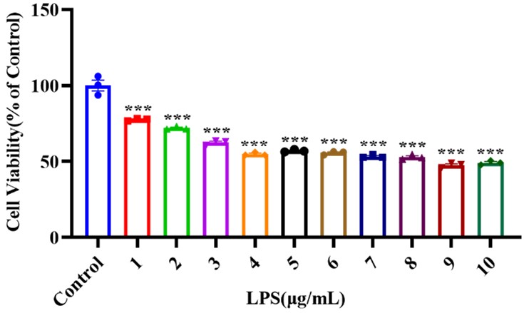
This model utilizes lipopolysaccharide (LPS) to activate murine BV2 microglia, simulating the secondary neuroinflammatory response that exacerbates ischemic brain damage. Concentration gradient screening was carried out via the CCK-8 cell viability assay, and 1 μg/mL LPS was selected as the optimal concentration for establishing the inflammatory model.

LPS concentration gradient screening in BV2 cells

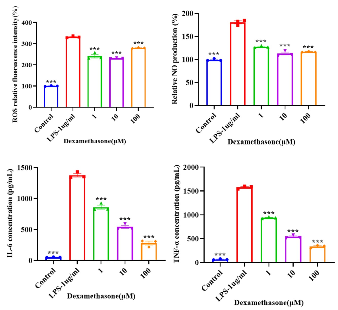
This model exhibited robust inflammatory responses, with increased ROS levels measured by fluorescence imaging, elevated NO production quantified via the Griess assay, and upregulated IL-6 and TNF-α secretion detected by ELISA. Dexamethasone significantly attenuated these pathological changes, with dose-dependent inhibition of IL-6 and TNF-α, validating the model as a reliable platform for anti-inflammatory drug screening.

Inflammatory responses in LPS-induced BV2 model

03Glutamate-Induced SH-SY5Y Cell Model

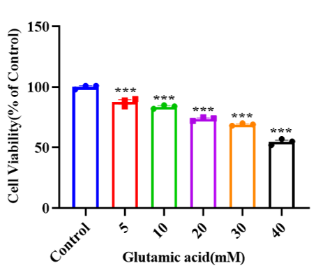
Glutamate excitotoxicity is a major driver of neuronal death during cerebral ischemia. This model uses glutamate to induce excitotoxic injury in SH-SY5Y neuroblastoma cells. Through concentration gradient screening via the CCK-8 cell viability assay, 20 mM glutamate was identified as the optimal modeling concentration. This condition stably induces neuronal injury and provides a robust platform for evaluating neuroprotective compounds against glutamate-induced excitotoxicity.

Glutamate concentration gradient screening in SH-SY5Y cells

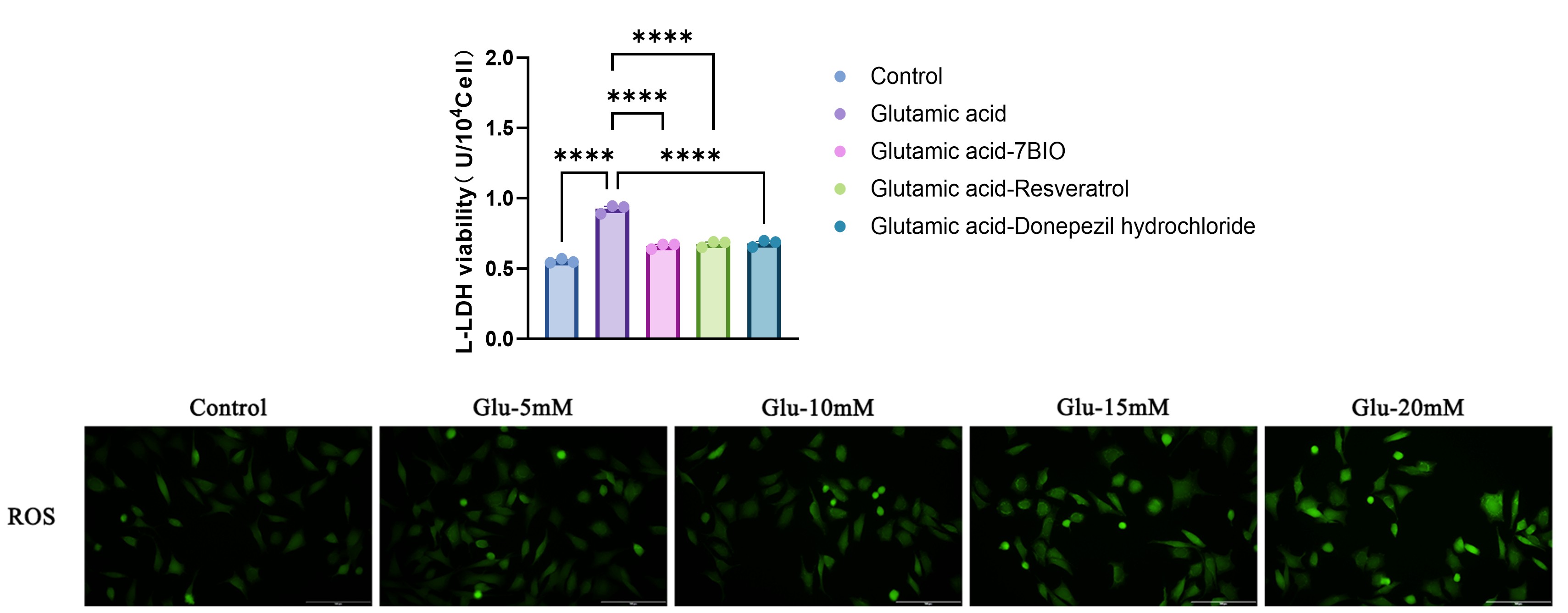
The glutamate-induced SH-SY5Y cell model exhibited robust excitotoxic responses, including increased oxidative stress by ROS detection and elevated LDH release by LDH cytotoxicity assay. Treatment with 7-BIO, Resveratrol, and Donepezil significantly attenuated glutamate-induced cytotoxicity, demonstrating the model's reliability and strong pharmacological responsiveness for excitotoxicity-targeted neuroprotection studies.

Excitotoxic responses in glutamate-induced SH-SY5Y model

04Glutamate-Induced Primary Hippocampal Neuron Injury Model

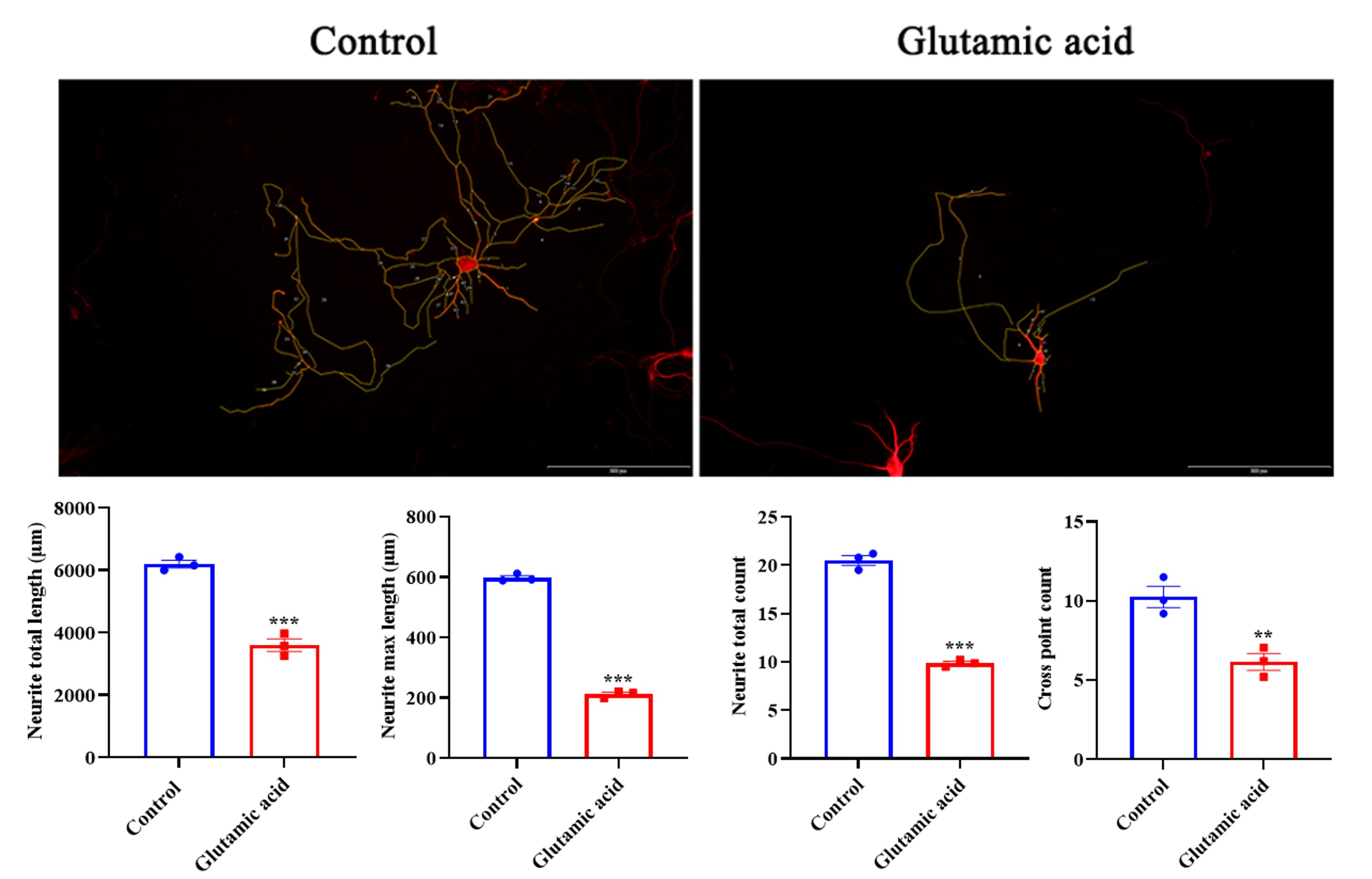
Building on the SH-SY5Y excitotoxicity model, this study extends to primary hippocampal neurons to capture more physiologically relevant morphological and structural responses to glutamate-induced injury.The glutamate-induced primary hippocampal neuron model was successfully established, characterized by significant decreases in total neurite length, maximum neurite length, neurite count, and branch points by neuronal morphological analysis. These findings reflect impaired neuronal morphology and complexity, providing a sensitive readout for structural neuroprotection and neuronal network integrity in the context of excitotoxic stroke injury.

Neuronal morphology in glutamate-induced primary hippocampal neuron model


Contact Us

We value your inquiries and are here to provide you with tailored solutions for your drug discovery and development needs. Whether you have questions, require more information, or are interested in discussing potential collaborations, our team of experts is just a message away.
Feel free to reach out to us.

We are a CRO service organization, not a hospital


Get a quote

Go top