
Our Payload Screening and Profiling service identifies and evaluates cytotoxic agents for antibody-drug conjugates (ADCs). We screen a broad range of payloads, including DNA-damaging agents, tubulin inhibitors, protein degraders, and immune modulators, to support efficacy and safety assessment for targeted cancer therapies.
Use assays such as Topoisomerase I-mediated DNA relaxation to evaluate how payloads biochemically disrupt essential cellular processes.
Use flow-based cell-cycle arrest assays, apoptosis assays, and high-content tubulin polymerization assays to analyze effects on cell division and cancer-cell death.
Evaluate single-payload activity and investigate synergistic effects from payload combinations to support therapeutic-strategy selection.
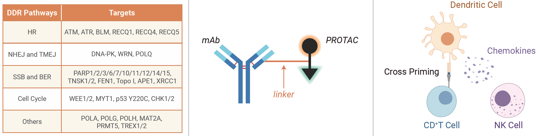
Explore Cancer Cell Panel Screening ↗Screen and evaluate DNA damage response inhibitors that may target cancer cells through their reliance on DNA-repair pathways.
Learn more ↗Assess targeted protein degradation payloads for challenging cancer targets that may otherwise be difficult to address.
Learn more ↗Evaluate STING agonists and STING-ISAC approaches designed to activate an immune response within the tumor microenvironment.
Topoisomerase I (Topo I) is a crucial enzyme in DNA replication and transcription. It alleviates torsional strain in the DNA helix by inducing single-strand breaks and re-ligation.
This assay assesses the ability of an ADC payload to inhibit Topo I and prevent DNA-strand re-ligation, helping determine payload potency and effectiveness as a Topo I inhibitor.
Contact us for detailed reference data ↗We offer absorbance-based and fluorescence-based tubulin polymerization assays to evaluate how ADC payloads disrupt microtubule dynamics and to provide mechanistic insight into their potential as anticancer agents.
Contact us for detailed reference data ↗Cell-cycle arrest assays evaluate how a cytotoxic payload affects cancer-cell proliferation by disrupting normal cell-cycle progression.
The resulting arrest can occur at phases including G1, S, or G2/M, depending on the payload mechanism of action, and can prevent cell division and contribute to cell death.
Contact us for detailed reference data ↗Flow-cytometry analysis quantifies apoptotic cells according to changes in cell size, complexity, and membrane asymmetry.
Annexin V FITC-A and PI-PE-A are used as apoptosis markers. Annexin V binds phosphatidylserine after its early-apoptosis translocation to the outer plasma membrane leaflet, while propidium iodide stains late apoptotic or dead cells but not live cells or early apoptotic cells.
Contact us for detailed reference data ↗We value your inquiries and are here to provide you with tailored solutions for your drug discovery and development needs. Whether you have questions, require more information, or are interested in discussing potential collaborations, our team of experts is just a message away.
Feel free to reach out to us.

Address: Bldg 16, Yd 18, Kechuang 13th St, Etown, Tongzhou Dist, Beijing, 100176, China
Email: marketing@ice-biosci.com
Tel:+86-10-67809840


Get a quote

Go top