
EN CN


Neuroscience

Return

In Vitro Parkinson's Disease Models & Assay Services

IN VITRO PARKINSON'S DISEASE MODELS

Parkinson's disease (PD) is the second most common neurodegenerative disorder, characterized by the progressive loss of dopaminergic neurons in the substantia nigra. Key pathological hallmarks include mitochondrial dysfunction, oxidative stress, and the accumulation of misfolded α-synuclein protein into Lewy bodies.

To accelerate therapeutic discovery and mechanistic understanding, in vitro cellular models have become indispensable tools in PD research. Our in vitro PD models recapitulate distinct disease-relevant pathways — ranging from oxidative stress and complex I inhibition to proteostasis impairment and α-synuclein aggregation — enabling compound screening, target validation, and mechanism-of-action (MoA) studies in a controlled, high-throughput environment prior to in vivo translation.

To comprehensively characterize these disease-relevant phenotypes, we offer a suite of integrated assay services, including CCK-8 cell viability assay, cellular ROS detection, Thioflavin T (ThT) assay, mitochondrial membrane potential assay (JC-1), and immunofluorescence — delivering robust, multi-dimensional readouts across cytotoxicity, oxidative stress, mitochondrial function, and pathological protein aggregation to empower data-driven decisions for your PD therapeutic pipeline.

MODEL SELECTION
Mechanism-driven in vitro model selection for Parkinson's disease drug discovery
Modeling MethodPrimary pathological mechanism modeledCommonly used cell typeKey molecular pathwaysUnique value
6-OHDA-inducedOxidative stress; effects on DAT/NET uptake; necrosis/apoptosisSH-SY5Y, PC12, primary neuronsROS, MAPK, Nrf2Oxidative stress testing
MPP⁺-inducedComplex I inhibition; DAT uptake; apoptosisSH-SY5Y, PC12, primary neuronsComplex I, mPTP, RTP801Mitochondrial drug screening
Rotenone-inducedComplex I inhibition; membrane permeable; apoptosis/autophagySH-SY5Y, primary neuronsComplex I, NLRP3, α-synEnvironmental toxin link; α-syn + mitochondrial
PFFs-inducedSeeded α-syn aggregation; autophagy/ER stressPrimary neurons, iPSC-DA neurons, SH-SY5YProteostasis, UPR, autophagyLewy body formation; anti-aggregation testing
α-Syn overexpression-inducedProteotoxic stress; apoptosis/autophagySH-SY5Y, PC12, primary neurons, iPSC-DA neuronsUPS, ALP, synaptic dysfunctionα-syn lowering; proteostasis testing
VALIDATED MODEL LIBRARY
Explore the model-specific validation workflow

Select a model below to review its biological rationale, experimental design, efficacy readouts, and representative data.

01Multi-PD Inducer-Induced SH-SY5Y & PC-12 Cell Models

In this model validation study, we systematically characterized in vitro Parkinson's disease models built on two widely adopted cell lines — SH-SY5Y and PC-12. Four classic pathological inducers (MPP⁺, rotenone, 6-OHDA, and α-synuclein PFFs) were used to trigger distinct PD-relevant pathogenic cascades. The models were profiled across three core pathological dimensions: cytotoxicity, α-synuclein accumulation and aggregation, and mitochondrial dysfunction coupled with oxidative stress. This multi-model, multi-readout approach enables direct comparison of complementary disease pathways for robust therapeutic screening.

Cytotoxicity evaluation in SH-SY5Y cells

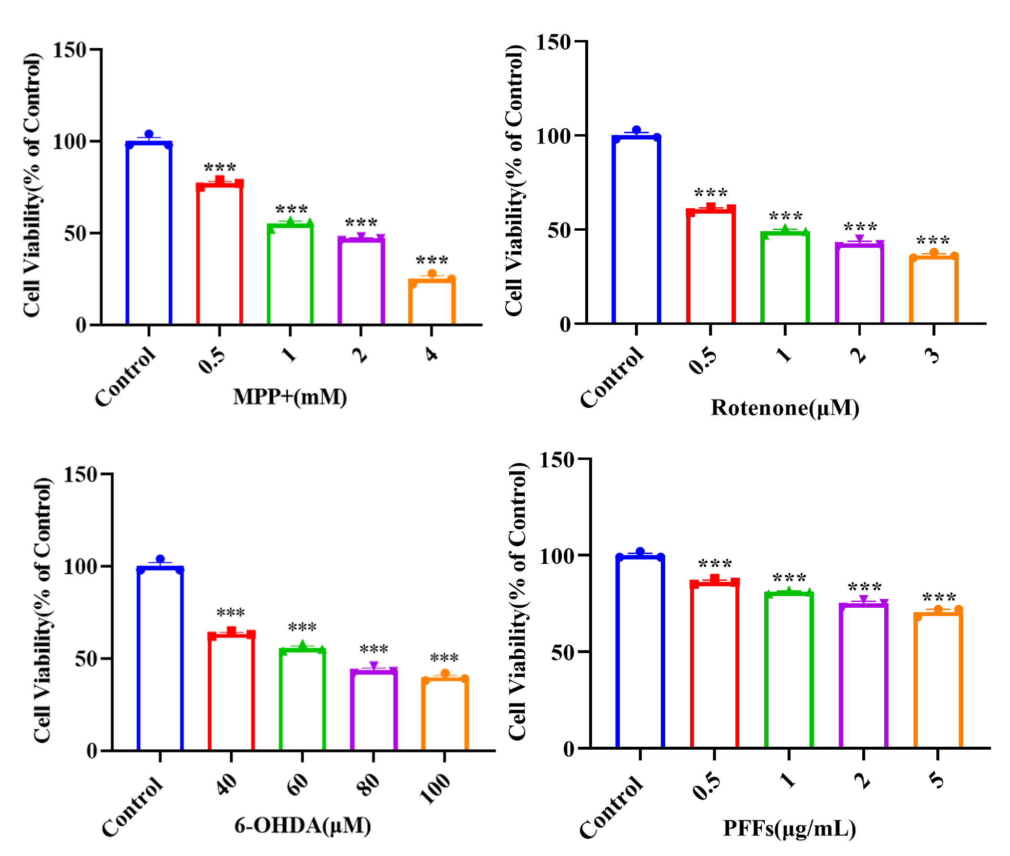
MPP⁺, rotenone, 6-OHDA, and PFFs each induced concentration-dependent loss of cell viability via CCK-8 cell viability assay in SH-SY5Y cells through distinct PD-relevant mechanisms, supporting their use as complementary in vitro models for compound screening and mechanistic evaluation.

Cytotoxicity evaluation of MPP+, rotenone, 6-OHDA, and PFFs in SH-SY5Y cells

α-synuclein accumulation and aggregation in SH-SY5Y & PC-12 cells

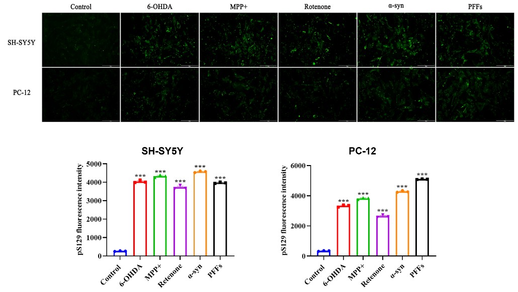
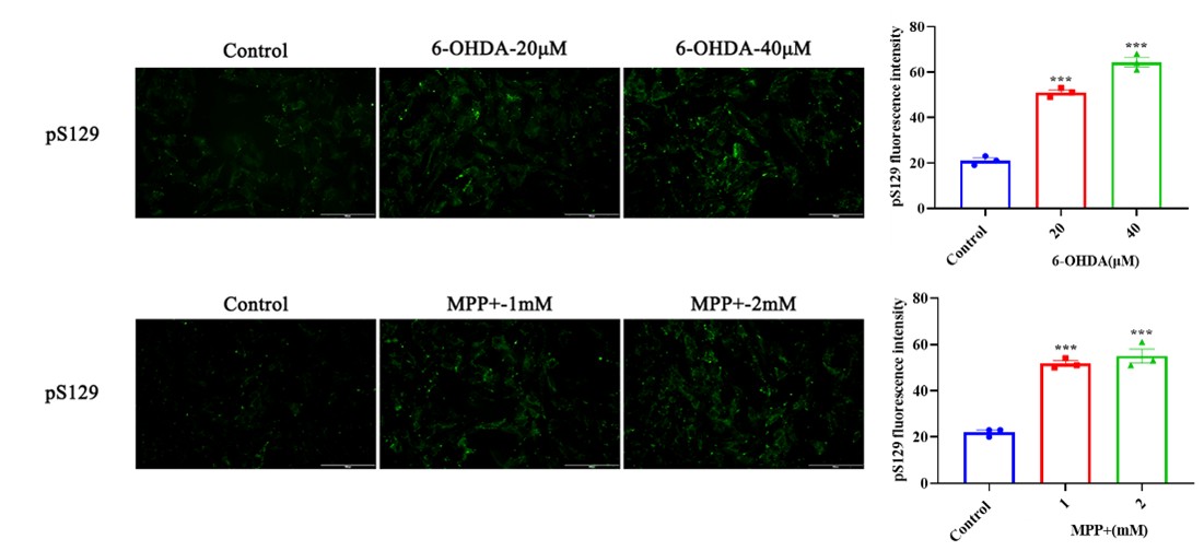
6-OHDA, MPP⁺, rotenone, α-synuclein, and PFFs each individually increased pSer129-α-synuclein fluorescence in SH-SY5Y and PC-12 cells, indicating activation of disease-relevant phosphorylation events.

pSer129-α-synuclein immunofluorescence in SH-SY5Y and PC-12 cells

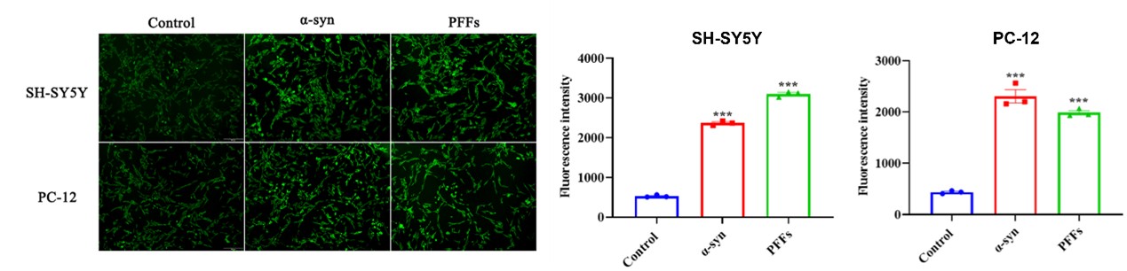
Furthermore, in α-synuclein- and PFFs-treated cells, increased ThT fluorescence by Thioflavin T (ThT) assay further confirmed enhanced α-synuclein fibril formation, supporting these in vitro models for evaluating α-synuclein-targeted therapeutic candidates.

Thioflavin T assay confirming α-synuclein fibril formation

Mitochondrial dysfunction and oxidative stress in SH-SY5Y cells

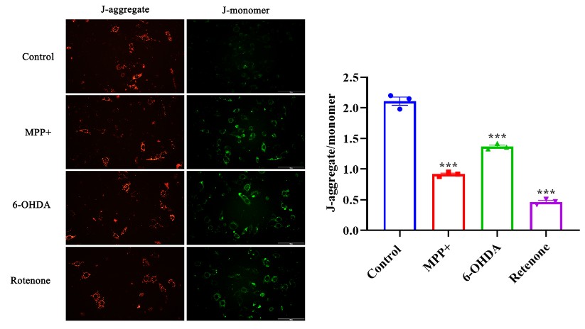
MPP⁺, 6-OHDA, and rotenone each significantly reduced the JC-1 aggregate-to-monomer ratio in the mitochondrial membrane potential assay (JC-1) in SH-SY5Y cells, demonstrating profound mitochondrial depolarization.

JC-1 mitochondrial membrane potential in SH-SY5Y cells

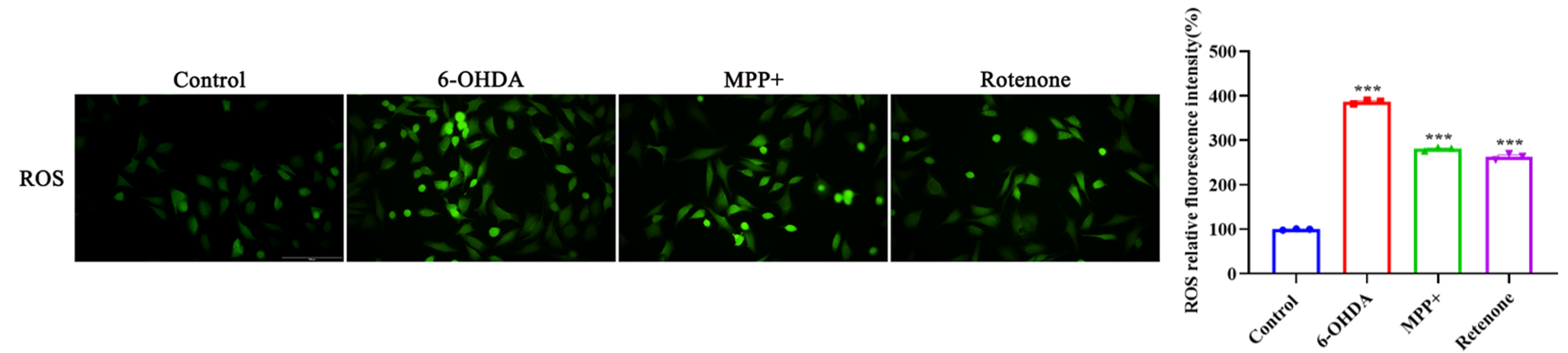
In addition, all three treatments showed elevated ROS fluorescence in the intracellular ROS detection assay, confirming oxidative stress as a shared and conserved pathological feature across mechanistically distinct PD cellular models.

Intracellular ROS detection in SH-SY5Y cells

02MPP⁺ & 6-OHDA-Induced Primary Midbrain Neuron Models

In this model validation study, we established toxin-induced Parkinson's disease models using primary midbrain neurons — a physiologically highly relevant cellular system that closely mimics the native dopaminergic neuronal context in vivo. Two well-characterized neurotoxins, MPP⁺ and 6-OHDA, were applied to trigger distinct PD-relevant pathogenic cascades, and two core pathological dimensions were systematically assessed: concentration-dependent cytotoxicity and pathological α-synuclein accumulation. This work delivers a biologically authentic in vitro platform for PD translational research and neuroprotective candidate evaluation.

Cytotoxicity evaluation in primary midbrain neurons

MPP⁺ and 6-OHDA each induced concentration-dependent loss of viability via CCK-8 cell viability assay in primary midbrain neurons, closely mimicking the selective dopaminergic neurodegeneration observed in vivo.

Cytotoxicity of MPP+ and 6-OHDA in primary midbrain neurons

α-synuclein accumulation in primary midbrain neurons

Both MPP⁺ and 6-OHDA treatments significantly increased pSer129-α-synuclein immunofluorescence, demonstrating that even acute mitochondrial dysfunction or oxidative stress can trigger pathological α-synuclein modifications in a physiologically relevant neuronal context.

pSer129-α-synuclein accumulation in primary midbrain neurons


Contact Us

We value your inquiries and are here to provide you with tailored solutions for your drug discovery and development needs. Whether you have questions, require more information, or are interested in discussing potential collaborations, our team of experts is just a message away.
Feel free to reach out to us.

We are a CRO service organization, not a hospital


Get a quote

Go top