
In vitro models of Major Depressive Disorder (MDD) serve as high-value translational tools in preclinical antidepressant discovery, bridging molecular target validation to distal biological efficacy within controlled cellular environments. These cellular platforms support high-throughput screening, simple operation, and relatively low assay cost, making them well suited for primary screening of large compound libraries. As a critical intermediate layer in the R&D pipeline, in vitro MDD models enable early-stage compound triage and mechanism-of-action deconvolution before in vivo studies. By prioritizing lead candidates with confirmed cellular efficacy and defined mechanisms, they can help reduce R&D spending, shorten development timelines, and improve the predictive reliability of subsequent in vivo efficacy evaluations.
| Modeling Method | Corresponding Depression Mechanism | Commonly Used Cell Types | Key Molecular Pathways | Main Phenotypic Characteristics | Unique Value in Efficacy Evaluation |
|---|---|---|---|---|---|
| Glucocorticoid-induced (Corticosterone/Dexamethasone) | HPA axis hyperactivity, impaired neuroplasticity | SH-SY5Y, PC12, primary cortical/hippocampal neurons | GR → inhibit BDNF transcription → block mTORC1 pathway | Cell survival rate ↓, apoptosis ↑, BDNF ↓, p-mTOR ↓, synaptic proteins (PSD-95/GluA1) ↓ | Most commonly used model, suitable for antidepressant screening and BDNF/mTOR pathway research |
| Glutamate-induced | Glutamate excitotoxicity | SH-SY5Y, PC12, primary cortical/hippocampal neurons | Overactivation of NMDA receptors → Ca2+ influx ↑ → ROS burst → mitochondrial damage | Cell survival rate ↓, calcium overload, ROS ↑, mitochondrial membrane potential ↓, antioxidant system (GSH) remodeling | Suitable for excitotoxicity research and rapid-acting antidepressant development |
| Lipopolysaccharide (LPS)-induced | Neuroinflammation hypothesis, abnormal activation of microglia | BV2 cells, microglia-neuron co-culture | TLR4/NF-κB pathway activation | Iba1 ↑ (microglial activation), IL-6/TNF-α/IL-1β ↑, indirectly leading to neuronal damage | Dedicated to neuroinflammatory mechanisms and inflammation-related depression research |
| Oxidative stress-induced (H2O2, 6-OHDA) | Mitochondrial dysfunction, oxidative damage | PC12, SH-SY5Y, primary cortical/hippocampal neurons | Lipid peroxidation chain reaction → DNA damage → mitochondrial membrane potential collapse | ROS ↑, MDA ↑, SOD ↓/GSH ↓, cell apoptosis rate ↑ | Suitable for oxidative response research and mitochondrial protection agent evaluation |
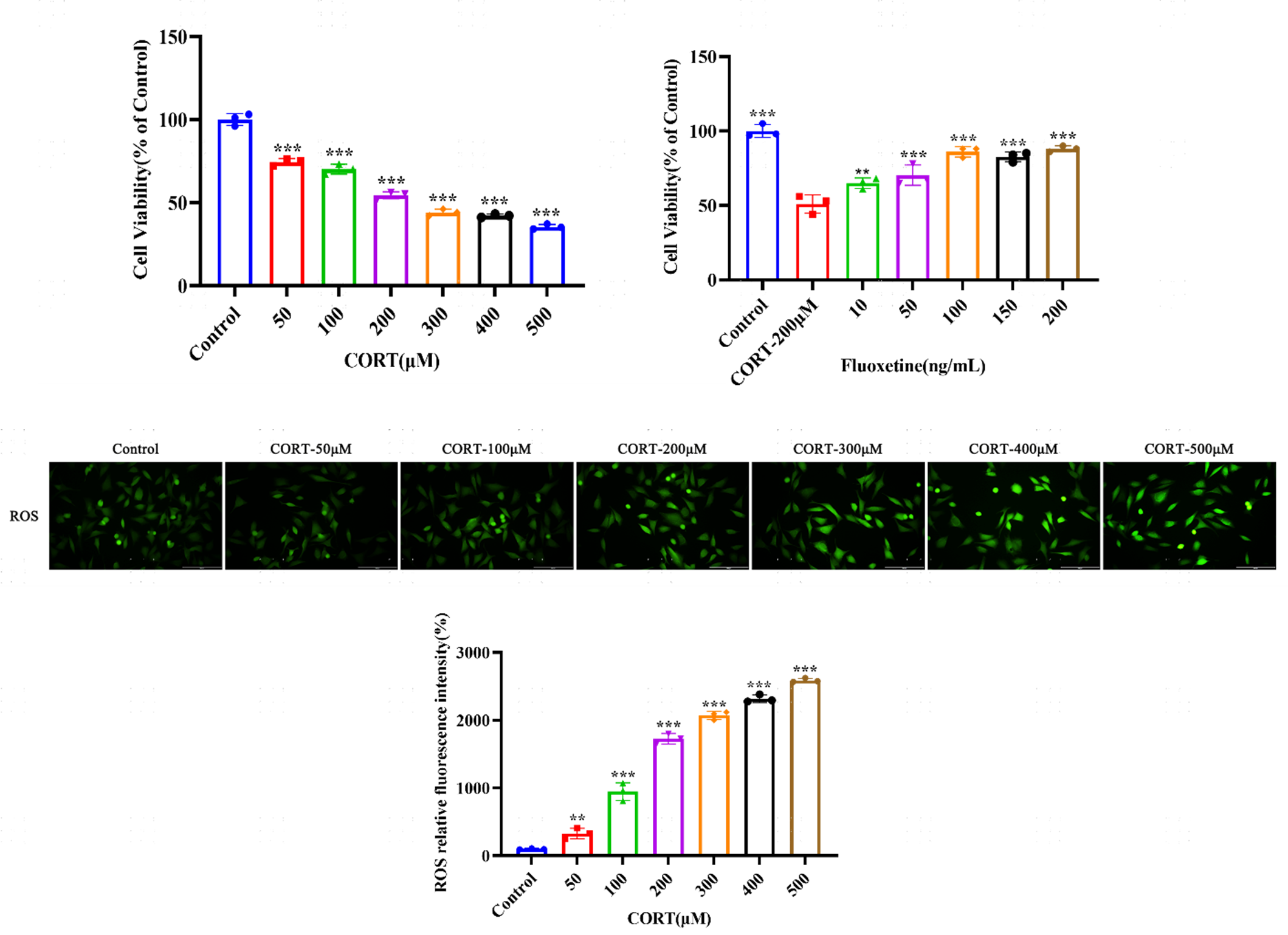
In the corticosterone-induced SH-SY5Y neuronal injury model, we offer CCK-8 cell viability assay and cellular ROS detection to evaluate neuroprotective effects of test compounds. Following co-incubation of SH-SY5Y cells with varying concentrations of corticosterone (CORT, 50–500 μM), cell viability decreases in a concentration-dependent manner, accompanied by a significant elevation in reactive oxygen species (ROS) levels. 200 μM CORT is selected as the optimal modeling concentration. Subsequent intervention with Fluoxetine (10–200 ng/mL) demonstrates a significant, concentration-dependent recovery in cell viability, indicating a clear neuroprotective effect.

Figure 1. CCK-8 cell viability assay and cellular ROS detection in CORT-induced SH-SY5Y injury model
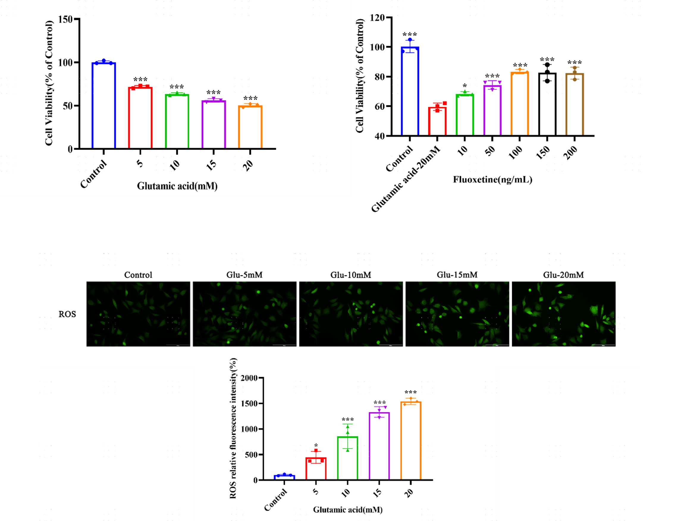
Co-incubation of SH-SY5Y cells with glutamate (Glu, 5–20 mM) results in a concentration-dependent reduction in cell viability alongside abnormal elevation of ROS levels. Using 20 mM Glu as the optimal modeling concentration, subsequent co-incubation with Fluoxetine (10–200 ng/mL) shows that the treatment effectively and dose-dependently reverses Glu-induced neurotoxicity and significantly enhances cell survival rate.

Figure 2. CCK-8 cell viability assay and cellular ROS detection in Glu-induced SH-SY5Y injury model
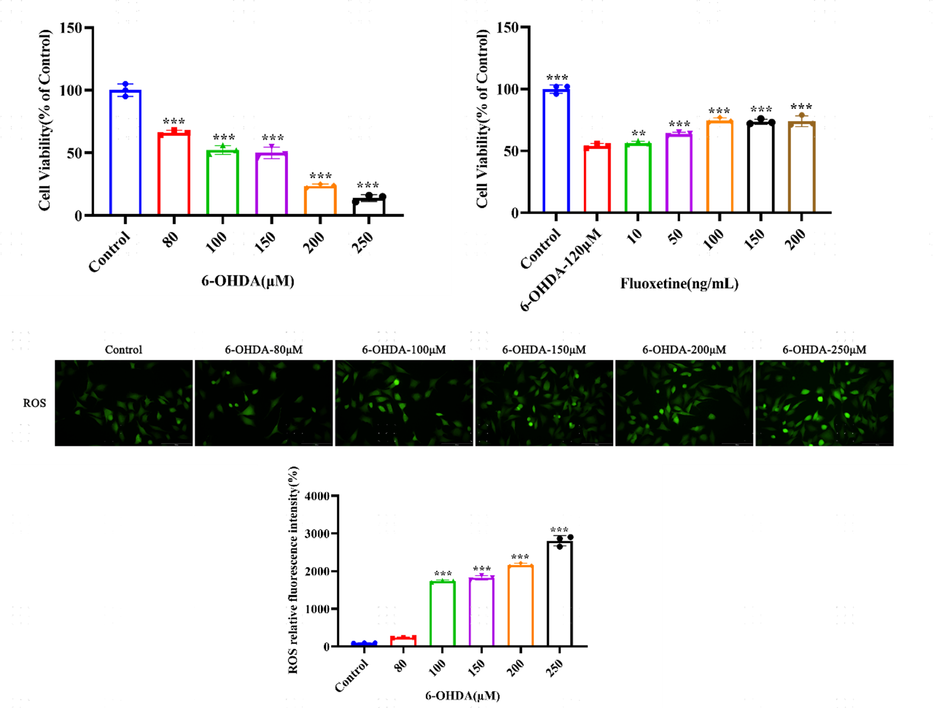
Treatment of SH-SY5Y cells with 6-hydroxydopamine (6-OHDA, 80–250 μM) leads to a concentration-dependent decline in cell viability and a simultaneous increase in intracellular ROS levels. The 120 μM 6-OHDA concentration is selected to establish the injury model. Following treatment with Fluoxetine (10–200 ng/mL), cell viability in 6-OHDA-damaged cells is effectively restored in a dose-dependent manner.

Figure 3. CCK-8 cell viability assay and cellular ROS detection in 6-OHDA-induced SH-SY5Y injury model
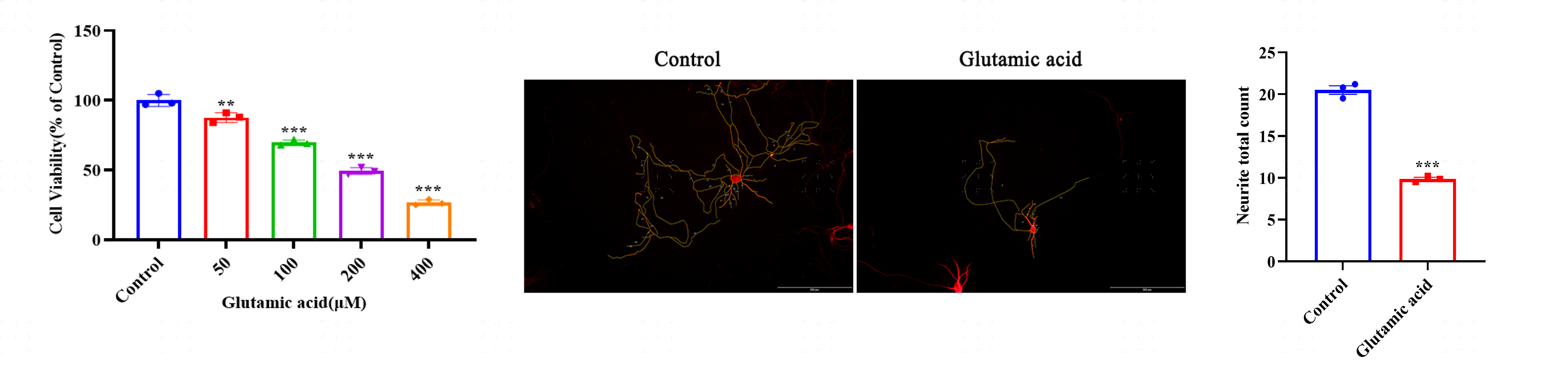
In the glutamate-induced primary cortical neuron model, we provide CCK-8 cell viability assay and neuronal morphological analysis to assess compound protective effects against glutamate excitotoxicity. Co-incubation of primary cortical neurons with Glu (50–400 μM) reveals a significant, concentration-dependent decrease in neuronal survival rate. Furthermore, morphological analysis indicates that Glu treatment significantly reduces neurite number in primary cortical neurons, confirming structural damage to the neuronal network.

Figure 4. CCK-8 viability assay and neuronal morphological analysis in Glu-induced primary cortical neuron model
We value your inquiries and are here to provide you with tailored solutions for your drug discovery and development needs. Whether you have questions, require more information, or are interested in discussing potential collaborations, our team of experts is just a message away.
Feel free to reach out to us.

Address: Bldg 16, Yd 18, Kechuang 13th St, Etown, Tongzhou Dist, Beijing, 100176, China
Email: marketing@ice-biosci.com
Tel:+86-10-67809840
Get a quote
Go top