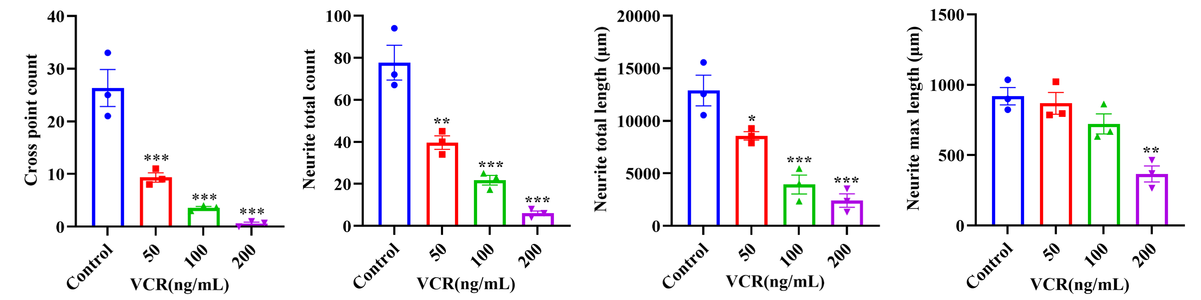

Pain is a complex sensory and biological response involving the peripheral and central nervous systems. Neuronal injury and dysfunction, particularly in sensory neurons, are key contributors to the development and persistence of neuropathic pain. Dorsal root ganglion (DRG) neurons play an essential role in sensing and transmitting peripheral pain signals. Damage to DRG neurons can lead to alterations in neuronal morphology, neurite integrity, and neuronal function, providing an important cellular basis for investigating pain-related mechanisms.
Among pain-related conditions, chemotherapy-induced peripheral neuropathy (CIPN) is one of the most common types of chronic pain encountered clinically. A range of chemotherapeutic agents, including vincristine, exert neurotoxicity that leads to DRG neuronal axonal injury and neurite degeneration, resulting in persistent pain in patients. In vitro DRG neuron models provide a controlled system for investigating chemotherapy-induced neuronal injury and pain-related phenotypes. By monitoring changes in neuronal morphology, neurite integrity, and molecular markers, these models enable detailed characterization of cellular responses to neurotoxic compounds and provide a useful platform for evaluating neuronal injury phenotypes.
At ICE Bioscience, we have established pain-related in vitro models using DRG neurons and integrated immunofluorescence (IF) imaging with quantitative morphological analysis to characterize chemotherapy-induced neuronal injury, delivering a robust, well-validated platform for neuroprotective drug screening and neuropathic pain research.
We offer a comprehensive pain ion channel panel covering core targets in pain signal transduction, including voltage-gated sodium/potassium/calcium channels, TRP channels, ligand-gated ion channels, acid-sensing ion channels and HCN channels. All assays are performed via manual patch clamp on human-derived targets, supporting both agonist and antagonist activity profiling for analgesic target validation and lead compound screening.
Pain Ion Channel Portfolio ↗Based on primary DRG neurons from multiple species (cynomolgus monkey, rat, mouse, etc.), we provide whole-cell patch-clamp electrophysiology testing. Focused on key pain targets such as TTX-resistant Nav1.8, the assays evaluate compound inhibitory potency under both resting and half-inactivated channel states. Native neuron assays deliver higher physiological relevance than recombinant cell line systems, bridging preclinical target screening and translational pain research.
DRG Neuron Electrophysiology ↗Review the biological rationale, experimental design, efficacy readouts, and representative data of the validated model below.
We value your inquiries and are here to provide you with tailored solutions for your drug discovery and development needs. Whether you have questions, require more information, or are interested in discussing potential collaborations, our team of experts is just a message away.
Feel free to reach out to us.

Address: Bldg 16, Yd 18, Kechuang 13th St, Etown, Tongzhou Dist, Beijing, 100176, China
Email: marketing@ice-biosci.com
Tel:+86-10-67809840


Get a quote

Go top